Categories:

Hyperventilation and Breathe-More Myth

Hyperventilation (definition) is breathing more than the medical norm.
http://www.normalbreathing.com/i-hyperventilation.php
Hyperventilation can be defined as breathing more than 10 liters of air per minute at rest for a 70-lg person.

Hyperventilation is very common these days. The large majority of people believe that it is worthwhile as well as healthy to breathe in more deeply (to hyperventilate) and obtain extra air in the respiratory system, medical doctors have the opposite view in regard to deep breathing or overbreathing.

The main initial result of overbreathing is lowered CO2 saturation of the arterial blood, and this reduces transport of O2 to cells. Let us consider how.

Hyperventilation does not improve blood oxygenation, which remains about the same, 98-99%, as during normal breathing. And physicians are right that hyperventilation does not raise O2 saturation of the arterial blood.

Hyperventilation reduces CO2 levels in the lungs and arterial blood. This effect takes place within 1-2 minutes.

Low arterial CO2 constricts arteries and arterioles reducing blood flow and O2 delivery to all vital organs. Dozens of studies confirmed this effect of hyperventilation.

Low CO2 also reduces O2 release in tissues. In other words, hyperventilation leads to the suppressed Bohr effect: less O2 is discharged from red blood cells due to reduced CO2 in tissues.

Nearly all chronic diseases are either based on or accompanied by hyperventilation. You can see these studies on NormalBreathing.com. Click the link at the top of this post.

Do not believe in “breathing-more” or “deep-breathing” myth.

Hundreds of doctors applied hyperventilation test in their medical practice. Let us consider how.

Numerous studies have made use of the overbreathing test (deliberate hyperventilation) when health care professionals required their clients to respire faster and heavier for 2 or 3 minutes so that these patients can reveal their signs and weakest organs of the human body. This kind of deep breathing test was particularly common with those specialists who taught breathing techniques. Multiple clinical research studies discovered that deep breathing causes many health problems.

Certainly, because a lot of individuals with diseases (usually over 90 percent) can recognize their signs during the deep breathing test, these individuals knew the root of their complications.

Awareness concerning regular respiration patterns helped the sufferers to find out the leading causes of their health symptoms. Now they can monitor their routine breathing so that they can notice too heavy breathing in every day lifestyle.

Initially, it happened to be discussed to the patient that deep breathing actions triggering over-breathing cause a decrease in carbon dioxide levels in the alveoli. These deep breathing actions consisted of coughing, yawning, sighing, sneezing as well as chest breathing. All are signs and symptoms of hyperventilation. So as to become healthy, those abnormal behaviors must be quit or avoided.

Learn more about hyperventilation symptoms: http://www.normalbreathing.com/hyperventilation-symptoms.php

Secondly, it was very important for the individual to recognize the features of upper body as well as abdominal respiration. Without a doubt, diaphragmatic breathing is usually represented by the observing adjectives: easy, quiet, slow, as well as light. On the flip side, chest respiration happens to be bulky, loud, too fast, as well as irregular.

Because psychological stress is a routine result to anxiety resulting in hyperventilation, one has the ability to end this. A person can slow down breathing. This reduces minute ventilation, lowers pulse rate, as well as boosts alveolar carbon dioxide concentrations. As a result, relaxation happens to be a useful therapeutic device in slashing deep breathing or hyperventilation as well as in improving one’s wellness as many studies related to various medical conditions have actually validated.

Furthermore, relaxation methods that slow down breathing can often be useful in getting rid of the negative effects of stress, deep breathing, and their subsequent uncommon physiological adjustments. In particular, the decrease of strain in the muscular tissues, particularly of the chest region, causes more calmness as well as better respiration.

URL of this YouTube video: https://www.youtube.com/watch?v=xhusT_X2e48

Find out more from Dr Artour Rakhimov’s website NormalBreathing.com
Posted in General.

Tagged with , , , , , , .


Sodium Deficiency and Stress

Also see:
Aldosterone, Sodium Deficiency, and Insulin Resistance
The Randle Cycle
Free Fatty Acids Suppress Cellular Respiration
Aldosterone as an endogenous cardiovascular toxin
Aldosterone and Thrombosis
Low Sodium Diet: High FFA, Insulin Resistance, Atherosclerosis
Sodium and Mortality – An Inverse Relationship
Elevated Levels of Sodium Blunt Response to Stress, Study Shows

“The absorption and retention of magnesium, sodium, and copper, and the synthesis of proteins, are usually poor in hypothyroidism. Salt craving is common in hypothyroidism, and eating additional sodium tends to raise the body temperature, and by decreasing the production of aldosterone, it helps to minimize the loss of magnesium, which in turn allows cells to respond better to the thyroid hormone. This is probably why a low sodium diet increases adrenalin production, and why eating enough sodium lowers adrenalin and improves sleep. The lowered adrenalin is also likely to improve intestinal motility.” -Ray Peat, PhD

J Hypertens. 1993 Dec;11(12):1381-6.
Effect of dietary salt restriction on urinary serotonin and 5-hydroxyindoleacetic acid excretion in man.
Sharma AM, Schorr U, Thiede HM, Distler A.
OBJECTIVE:
To determine the effect of dietary salt restriction on urinary excretion of serotonin and its principal metabolite 5-hydroxyindoleacetic acid (5-HIAA) in man.
DESIGN:
We studied 16 healthy male volunteers (age range 20-28 years) who ate a standard diet containing 20 mmol/day NaCl, to which either 220 mmol/day NaCl or placebo was added as a supplement for 1 week each, according to a randomized, single-blind crossover design.
METHODS:
Urinary excretion of serotonin, 5-HIAA, noradrenaline and vanillylmandelic acid (VMA) were measured during the low- and high-salt periods using reverse-phase high-performance liquid chromatography.
RESULTS:
During the low-salt diet, 24-h urinary excretion of serotonin increased by 42%, accompanied by a 52% rise in the excretion of 5-HIAA. Salt restriction also increased noradrenaline excretion by 77% and VMA excretion by 40%. Regression analysis revealed a strong positive relationship between the excretion of serotonin and of noradrenaline (r = 0.84, P < 0.001) and between that of 5-HIAA and of VMA (r = 0.74, P < 0.001).
CONCLUSIONS:
Salt restriction stimulates the serotonergic system in man. Stimulation of this system, in conjunction with the sympathetic nervous system, may contribute to renal sodium conservation during dietary salt restriction in man.

J Clin Endocrinol Metab. 1991 Nov;73(5):975-81.
Effects of sodium supplementation during energy restriction on plasma norepinephrine levels in obese women.
Gougeon R, Mitchell TH, Larivière F, Abraham G, Montambault M, Marliss EB.
We tested whether sodium restriction would counteract the decrease in sympathetic nervous system activity usually associated with marked energy restriction. The effects of two levels of energy restriction, with different sodium intakes, on plasma norepinephrine (NE) levels while supine and in response to standing were studied. Twenty-two healthy normotensive obese female subjects (body mass index, 34 +/- 1 kg/m2; weight, 90 +/- 2 kg) followed one of three 3-week protocols: 1) total fasting with 80 mmol/day NaCl, 2) a very low energy diet (VLED) containing 1.7 MJ, 93 g protein, and 90 mmol Na/day, with an additional 60 mmol/day NaCl supplement, or 3) total fasting without NaCl (0 Na fast). At the end of the baseline isocaloric diet and of total fasts or VLED, pulse, blood pressure, and plasma NE were measured after 4 h of recumbency and 5 and 10 min after assuming the upright posture. These measurements were repeated after 1 L physiological saline was infused into the 0 Na fast subjects. Cumulative negative sodium balance was observed only in the 0 Na fasting subjects. Supine blood pressure decreased from baseline with fasting, but not with the VLED. The decreases in systolic pressure and increases in heart rate on standing observed with all diets were greatest with the 0 Na fast. Supine plasma NE (vs. baseline value) declined (P less than 0.05) with the VLED, remained unchanged with the Na supplemented fast, but increased with the 0 Na fast (P less than 0.05). The upright plasma NE values were highest in the 0 Na fast subjects, but lower after the saline infusion as well as in the subjects on the VLED. Thus, the decrease in NE due to energy restriction with normal sodium intake was counteracted by moderate sodium restriction, and levels increased with zero sodium intake. Therefore, sodium depletion can override the suppressive effect of energy restriction and, instead, increase the activity of the sympathetic nervous system, as reflected by plasma NE.

J Card Fail. 2009 Dec;15(10):864-73.
Long-term effects of dietary sodium intake on cytokines and neurohormonal activation in patients with recently compensated congestive heart failure.
Parrinello G, Di Pasquale P, Licata G, Torres D, Giammanco M, Fasullo S, Mezzero M, Paterna S.
BACKGROUND:
A growing body of evidence suggests that the fluid accumulation plays a key role in the pathophysiology of heart failure (HF) and that the inflammatory and neurohormonal activation contribute strongly to the progression of this disorder.
METHODS AND RESULTS:
The study evaluated the long-term effects of 2 different sodium diets on cytokines neurohormones, body hydration and clinical outcome in compensated HF outpatients (New York Heart Association Class II). A total of 173 patients (105 males, mean age 72.5+/-7) recently hospitalized for worsening advanced HF and discharged in normal hydration and in clinical compensation were randomized in 2 groups (double blind). In Group 1, 86 patients received a moderate restriction in sodium (120mmol to 2.8g/day) plus oral furosemide (125 to 250mg bid); in Group 2, 87 patients: received a low-sodium diet (80mmol to 1.8g/day) plus oral furosemide (125 to 250mg bid). Both groups were followed for 12 months and the treatment was associated with a drink intake of 1000mL daily. Neurohormonal (brain natriuretic peptide, aldosterone, plasma rennin activity) and cytokines values (tumor necrosis factor-alpha, interleukin-6) were significantly reduced with a significant increase of the anti-inflammatory cytokine interleukin-10 at 12 months in normal, P < .0001) than low-sodium group. The low-sodium diet showed a significant activation of neurohormones and cytokines and worsening the body hydration, whereas moderate sodium restriction maintained dry weigh and improved outcome in the long term.
CONCLUSIONS:
Our results appear to suggest a surprising efficacy of a new strategy to improve the chronic diuretic response by increasing Na intake and limiting fluid intake. This counterintuitive approach underlines the need for a better understanding of factors that regulate sodium and water handling in chronic congestive HF. A larger sample of patients and further studies are required to evaluate whether this is due to the high dose of diuretic used or the low-sodium diet.

Posted in General.

Tagged with , , , , , , , , , , , , , , , , .


Low Sodium Diet: High FFA, Insulin Resistance, Atherosclerosis

Also see:
Aldosterone, Sodium Deficiency, and Insulin Resistance
The Randle Cycle
Free Fatty Acids Suppress Cellular Respiration
Aldosterone as an endogenous cardiovascular toxin
Aldosterone and Thrombosis
Sodium Deficiency and Stress

J Endocrinol. 2005 Jun;185(3):429-37.
Low salt intake modulates insulin signaling, JNK activity and IRS-1ser307 phosphorylation in rat tissues.
Prada PO, Coelho MS, Zecchin HG, Dolnikoff MS, Gasparetti AL, Furukawa LN, Saad MJ, Heimann JC.
A severe restriction of sodium chloride intake has been associated with insulin resistance and obesity. The molecular mechanisms by which the low salt diet (LS) can induce insulin resistance have not yet been established. The c-jun N-terminal kinase (JNK) activity has been involved in the pathophysiology of obesity and induces insulin resistance by increasing inhibitory IRS-1(ser307) phosphorylation. In this study we have evaluated the regulation of insulin signaling, JNK activation and IRS-1(ser307) phophorylation in liver, muscle and adipose tissue by immunoprecipitation and immunoblotting in rats fed with LS or normal salt diet (NS) during 9 weeks. LS increased body weight, visceral adiposity, blood glucose and plasma insulin levels, induced insulin resistance and did not change blood pressure. In LS rats a decrease in PI3-K/Akt was observed in liver and muscle and an increase in this pathway was seen in adipose tissue. JNK activity and IRS-1(ser307) phosphorylation were higher in insulin-resistant tissues. In summary, the insulin resistance, induced by LS, is tissue-specific and is accompanied by activation of JNK and IRS-1(ser307) phosphorylation. The impairment of the insulin signaling in these tissues, but not in adipose tissue, may lead to increased adiposity and insulin resistance in LS rats.

Physiol Res. 2000;49(2):197-205.
Low-salt diet alters the phospholipid composition of rat colonocytes.
Mrnka L, Nováková O, Novák F, Tvrzická E, Pácha J.
The effect of low-salt diet on phospholipid composition and remodeling was examined in rat colon which represents a mineralocorticoid target tissue. To elucidate this question, male Wistar rats were fed a low-salt diet and drank distilled water (LS, low-salt group) or saline instead of water (HS, high-salt group) for 12 days before the phospholipid concentration and fatty acid composition of isolated colonocytes were examined. The dietary regimens significantly influenced the plasma concentration of aldosterone which was high in LS group and almost zero in HS group. Plasma concentration of corticosterone was unchanged. When expressed in terms of cellular protein content, a significantly higher concentration of phospholipids was found in LS group, with the exception of sphingomyelin (SM) and phosphatidylserine (PS). Phosphatidylcholine (PC) and phosphatidylethanolamine (PE) accounted for more than 70% of total phospholipids in both groups. A comparison of phospholipid distribution in LS and HS groups demonstrated a higher percentage of PE and a small, but significant, decrease of PC and SM in LS group. The percentage of phosphatidylinositol (PI), PS and cardiolipin (CL) were not affected by mineralocorticoid treatment. With respect to the major phospholipids (PE, PC), a higher level of n-6 polyunsaturated fatty acids (PUFA) and lower levels of monounsaturated fatty acids were detected in PC of LS group. The increase of PUFA predominantly reflected an increase in arachidonic acid by 53%. In comparison to the HS group, oleic acid content was decreased in PC and PE isolated from colonocytes of the LS group. Our data indicate that alterations in phospholipid concentration and metabolism can be detected in rats with secondary hyperaldosteronism. The changes in phospholipid concentration and their fatty acid composition during fully developed effect of low dietary Na+ intake may reflect a physiologically important phenomenon with long-term consequences for membrane structure and function.

J Lipid Res. 2003 Apr;44(4):727-32. Epub 2003 Jan 16.
Dietary sodium chloride restriction enhances aortic wall lipid storage and raises plasma lipid concentration in LDL receptor knockout mice.
Catanozi S, Rocha JC, Passarelli M, Guzzo ML, Alves C, Furukawa LN, Nunes VS, Nakandakare ER, Heimann JC, Quintão EC.
This study aimed at measuring the influence of a low salt diet on the development of experimental atherosclerosis in moderately hyperlipidemic mice. Experiments were carried out on LDL receptor (LDLR) knockout (KO) mice, or apolipoprotein E (apoE) KO mice on a low sodium chloride diet (LSD) as compared with a normal salt diet (NSD). On LSD, the rise of the plasma concentrations of TG and nonesterified fatty acid (NEFA) was, respectively, 19% and 34% in LDLR KO mice, and 21% and 35% in apoE KO mice, and that of plasma cholesterol was limited to the LDLR KO group alone (15%). Probably due to the apoE KO severe hypercholesterolemia, the arterial inner-wall fat storage was not influenced by the diet salt content and was far more abundant in the apoE KO than in the LDLR KO mice. However, in the less severe hypercholesterolemia of the LDLR KO mice, lipid deposits on the LSD were greater than on the NSD. Arterial fat storage correlated with NEFA concentrations in the LDLR KO mice alone (n = 14, P = 0.0065). Thus, dietary sodium chloride restriction enhances aortic wall lipid storage in moderately hyperlipidemic mice.

Metabolism. 2011 Jul;60(7):965-8. Epub 2010 Oct 30.
Low-salt diet increases insulin resistance in healthy subjects.
Garg R, Williams GH, Hurwitz S, Brown NJ, Hopkins PN, Adler GK.
Low-salt (LS) diet activates the renin-angiotensin-aldosterone and sympathetic nervous systems, both of which can increase insulin resistance (IR). We investigated the hypothesis that LS diet is associated with an increase in IR in healthy subjects. Healthy individuals were studied after 7 days of LS diet (urine sodium <20 mmol/d) and 7 days of high-salt (HS) diet (urine sodium >150 mmol/d) in a random order. Insulin resistance was measured after each diet and compared statistically, unadjusted and adjusted for important covariates. One hundred fifty-two healthy men and women, aged 39.1 ± 12.5 years (range, 18-65) and with body mass index of 25.3 ± 4.0 kg/m(2), were included in this study. Mean (SD) homeostasis model assessment index was significantly higher on LS compared with HS diet (2.8 ± 1.6 vs 2.4 ± 1.7, P < .01). Serum aldosterone (21.0 ± 14.3 vs 3.4 ± 1.5 ng/dL, P < .001), 24-hour urine aldosterone (63.0 ± 34.0 vs 9.5 ± 6.5 μg/d, P < .001), and 24-hour urine norepinephrine excretion (78.0 ± 36.7 vs 67.9 ± 39.8 μg/d, P < .05) were higher on LS diet compared with HS diet. Low-salt diet was significantly associated with higher homeostasis model assessment index independent of age, sex, blood pressure, body mass index, serum sodium and potassium, serum angiotensin II, plasma renin activity, serum and urine aldosterone, and urine epinephrine and norepinephrine. Low-salt diet is associated with an increase in IR. The impact of our findings on the pathogenesis of diabetes and cardiovascular disease needs further investigation.

Posted in General.

Tagged with , , , , , , , , , .


Syphillis as Aids

Syphilis as Aids

Posted in General.

Tagged with , .


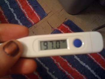
Inflammation from Decrease in Body Temperature

Also see:
Melatonin Lowers Body Temperature
Menopausal Estrogen Therapy Lowers Body Temperature
Temperature and Pulse Basics & Monthly Log
Thyroid, Temperature, Pulse
Ray Peat, PhD on Thyroid, Temperature, Pulse, and TSH

“A slight decrease in temperature can promote inflammation (Matsui, et al., 2006). The thermogenic substances–dietary protein, sodium, sucrose, thyroid and progesterone–are antiinflammatory for many reasons, but very likely the increased temperature itself is important.” -Ray Peat, PhD

“For example, keeping cells in a well oxygenated state with thyroid hormone and carbon dioxide will shift the balance from estradiol toward the weaker estrone. The thyroid stimulation will cause the liver to excrete estrogen more quickly, and will help to prevent the formation of aromatase in the tissues. Low temperature is one of the factors that increases the formation of estrogen. Lactic acid, serotonin, nitric oxide, prostaglandins, and the endorphins will be decreased by the shift toward efficient oxidative metabolism.” -Ray Peat, PhD

J Neurosurg Anesthesiol. 2006 Jul;18(3):189-93.
Mild hypothermia promotes pro-inflammatory cytokine production in monocytes.
Matsui T, Ishikawa T, Takeuchi H, Okabayashi K, Maekawa T.
Hypothermia is often associated with compromised host defenses and infection. Deteriorations of immune functions related to hypothermia have been investigated, but the involvement of cytokines in host defense mechanisms and in infection remains unclear. We have previously shown that mild hypothermia modifies cytokine production by peripheral blood mononuclear cells. In this study, the effects of hypothermia on the monocytic production of several cytokines and nitric oxide (NO) were determined. Monocytes obtained from 10 healthy humans were cultured with lipopolysaccharide (LPS) under hypothermic (33 degrees C) or normothermic (37 degrees C) conditions for 48 hours. We performed flow cytometric analysis for simultaneous measurement of interleukin (IL)-8, IL-1beta, IL-6, IL-10, IL-12p70, and tumor necrosis factor (TNF)-alpha in culture supernatants. NO production was quantified as accumulation of nitrite in the medium by a colorimetric assay. Compared with normothermia, mild hypothermia raised the levels of IL-1beta, IL-6, IL-12p70, and TNF-alpha produced by monocytes stimulated with LPS. On calculating the ratios of these elevated cytokines to IL-10, however, only IL-12p70/IL-10 and TNF-alpha/IL-10 ratios were significantly elevated under hypothermic conditions. In contrast, hypothermia did not affect NO production. This study demonstrates that mild hypothermia affects the balance of cytokines produced by monocytes, leading to a pro-inflammatory state. Specifically, monocytic IL-12 and TNF-alpha appear to be involved in the immune alterations observed in mild hypothermia. However, the clinical significance of these phenomena remains to be clarified.

Posted in General.

Tagged with , , , , , , , , , , .


Topical Vitamin E and ultraviolet radiation on human skin

Also see:
Sunburn, PUFA, Prostaglandins, and Aspirin
Unsaturated Fats and Age Pigment
PUFA Accumulation & Aging
Niacinamide and the Skin

Med Cutan Ibero Lat Am. 1990;18(4):269-72.
[Topical Vitamin E and ultraviolet radiation on human skin].
[Article in Spanish]
Rampoldi R, Macedo N, Alallon W, Sanguimetti J.
The point is the relation between the tissue damage caused by UV radiations, the production of free radicals (lipoperoxidation) and the particular action of vitamin E on human skin. Through histopathologic changes and malondialdehyde dosification results, are analyzed, therefore a close relation between UV radiation, free radicals, lipoperoxidation, and tissue damage, is proved on human skin. Furthermore the protective action of topic vitamin E antioxidant which diminishes the lipoperoxidation and the tissue damage is apparent.

================================
PUFA wastes Vitamin E

Int J Vitam Nutr Res. 2000 Mar;70(2):31-42.
Relationship between vitamin E requirement and polyunsaturated fatty acid intake in man: a review.
Valk EE1, Hornstra G.
Vitamin E is the general term for all tocopherols and tocotrienols, of which alpha-tocopherol is the natural and biologically most active form. Although gamma-tocopherol makes a significant contribution to the vitamin E CONTENT in foods, it is less effective in animal and human tissues, where alpha-tocopherol is the most effective chain-breaking lipid-soluble antioxidant. The antioxidant function of vitamin E is critical for the prevention of oxidation of tissue PUFA. Animal experiments have shown that increasing the degree of dietary fatty acid unsaturation increases the peroxidizability of the lipids and reduces the time required to develop symptoms of vitamin E deficiency. From these experiments, relative amounts of vitamin E required to protect the various fatty acids from being peroxidized, could be estimated. Since systematic studies on the vitamin E requirement in relation to PUFA consumption have not been performed in man, recommendations for vitamin E intake are based on animal experiments and human food intake data. An intake of 0.6 mg alpha-tocopherol equivalents per gram linoleic acid is generally seen as adequate for human adults. The minimum vitamin E requirement at consumption of fatty acids with a higher degree of unsaturation can be calculated by a formula, which takes into account the peroxidizability of unsaturated fatty acids and is based on the results of animal experiments. There are, however, no clear data on the vitamin E requirement of humans consuming the more unsaturated fatty acids as for instance EPA (20:5, n-3) and DHA (22:6, n-3). Studies investigating the effects of EPA and DHA supplementation have shown an increase in lipid peroxidation, although amounts of vitamin E were present that are considered adequate in relation to the calculated oxidative potential of these fatty acids. Furthermore, a calculation of the vitamin E requirement, using recent nutritional intake data, shows that a reduction in total fat intake with a concomitant increase in PUFA consumption, including EPA and DHA, will result in an increased amount of vitamin E required. In addition, the methods used in previous studies investigating vitamin E requirement and PUFA consumption (for instance erythrocyte hemolysis), and the techniques used to assess lipid peroxidation (e.g. MDA analysis), may be unsuitable to establish a quantitative relation between vitamin E intake and consumption of highly unsaturated fatty acids. Therefore, further studies are required to establish the vitamin E requirement when the intake of longer-chain, more-unsaturated fatty acids is increased. For this purpose it is necessary to use functional techniques based on the measurement of lipid peroxidation in vivo. Until these data are available, the widely used ratio of at least 0.6 mg alpha-TE/g PUFA is suggested. Higher levels may be necessary, however, for fats that are rich in fatty acids containing more than two double bonds.

Br J Nutr. 2015 Oct 28;114(8):1113-22. doi: 10.1017/S000711451500272X. Epub 2015 Aug 21.
Vitamin E function and requirements in relation to PUFA.
Raederstorff D1, Wyss A1, Calder PC2, Weber P1, Eggersdorfer M1.
Vitamin E (α-tocopherol) is recognised as a key essential lipophilic antioxidant in humans protecting lipoproteins, PUFA, cellular and intra-cellular membranes from damage. The aim of this review was to evaluate the relevant published data about vitamin E requirements in relation to dietary PUFA intake. Evidence in animals and humans indicates a minimal basal requirement of 4-5 mg/d of RRR-α-tocopherol when the diet is very low in PUFA. The vitamin E requirement will increase with an increase in PUFA consumption and with the degree of unsaturation of the PUFA in the diet. The vitamin E requirement related to dietary linoleic acid, which is globally the major dietary PUFA in humans, was calculated to be 0·4-0·6 mg of RRR-α-tocopherol/g of linoleic acid. Animal studies show that for fatty acids with a higher degree of unsaturation, the vitamin E requirement increases almost linearly with the degree of unsaturation of the PUFA in the relative ratios of 0·3, 2, 3, 4, 5 and 6 for mono-, di-, tri-, tetra-, penta- and hexaenoic fatty acids, respectively. Assuming a typical intake of dietary PUFA, a vitamin E requirement ranging from 12 to 20 mg of RRR-α-tocopherol/d can be calculated. A number of guidelines recommend to increase PUFA intake as they have well-established health benefits. It will be prudent to assure an adequate vitamin E intake to match the increased PUFA intake, especially as vitamin E intake is already below recommendations in many populations worldwide.

Posted in General.

Tagged with , , , , , , , , , , , , , , .


Multiple autoimmune syndrome

Also see:
Autoimmune Disease and Estrogen Connection

J Pak Med Assoc. 2010 Oct;60(10):863-5.
Multiple autoimmune syndrome: Hashimoto’s thyroiditis, coeliac disease and systemic lupus erythematosus (SLE).
Latif S, Jamal A, Memon I, Yasmeen S, Tresa V, Shaikh S.
Various autoimmune diseases have association with each other but it is very rare to see multiple autoimmune diseases in one patient. Presence of more than two autoimmune diseases in one patient is known as multiple autoimmune syndrome (MAS). We report the case of an 11 years old girl who presented with history of swelling in front of the neck along with constipation, anorexia, weight gain and increasing pallor over a period of six months. Additionally she had an episodic history of joint pains and abdominal pain with no specific relation to diet, time, other gastrointestinal or genitourinary symptom. Hypothyroid goiter (Autoimmune thyroiditis, Hashimoto’s thyroidits) was diagnosed by raised thyroid stimulating hormone (TSH), low T4 and presence of thyroid specific antibodies in blood. Patient was discharged on tablet Levothyroxine to which she responded well with reduction in size of the swelling and relief of the symptoms except for the joint pains and abdominal pain. To evaluate the persistent symptoms she was investigated further for other autoimmune diseases and was diagnosed to be having systemic lupus erythematosus (SLE) and Coeliac disease also. The final diagnosis was multiple autoimmune syndrome (Hashimoto’s thyroiditis, Coeliac disease and SLE).

Posted in General.

Tagged with .


180 Nutrition Program Testimonial – Complete Wellness

Female, California, Age 40

The top things I achieved in Functional Performance Systems ‘180 Nutrition Program’ are:

Stress management: Tremendous improvement in mental clarity and physical relaxation and calm on a deep level. My body feels amazing every day; I can literally just sit here and feel physically blissful. I don’t think I’ve ever felt so relaxed in my life. I’m able to psychologically handle stress from a much more calm mental space. I’m able to be more productive in every area of my life and therefore have opened myself up to better opportunities and experiences. Through applying 180 nutritional guidelines and principles, I’m experiencing a marked improvement in the quality of my existence!

Digestion: I’ve healed the lining of gut on a deep level without the dependence on supplements, which is incredible for me. I feel like my digestive system is running like a machine, absorbing and utilizing the necessary nutrients and efficiently eliminating. I feel light and energetic.

Sexual health: Although very personal and I debated whether commenting on it, optimal sexual health is a part of our lives as human beings. The hormonal balance I’ve achieved in this program through detoxifying excess estrogen as well as inhibiting an estrogen stress response (among other hormonal factors) has led to increased orgasmic capacity. This is not only rewarding on a personal physical level, emotionally and mentally, but will lend to positive intimate shared experience with a partner.

Body composition: I’ve followed a no fruit/low to no sugar, low fat nutrition philosophy for the last 18 years, worked out with weights and incorporated a lot of cardiovascular exercise ~ all of this a set-up for a ruined metabolism and low thyroid function. I’ve always kept my body fat somewhere between 17-19% which looks fit and healthy, but looks are very deceiving! Over the past six months I’ve been eating tons of fruit, tons of saturated fat, laying completely off of any cardio, minimizing my workouts to one session of sprints or squats every 1-2 weeks, and allowing my body to rest and heal. I started out at 111 lbs, appx.19% body fat, and I am now 120 lbs and appx. 21% body fat, so I gained 9 pounds and only 1-2% body fat. To gain that much lean body mass and that little of body fat by incorporating healing foods and rest is incredible. I am literally getting daily compliments on my figure from strangers. I feel so much more feminine and love the weight distribution. This is all due to the positive hormonal response elicited from the food in this program, and proper rest and recovery.

Mood: I feel SO happy every day due to my balanced hormone levels and well functioning body systems! When I started this program I was extremely stressed and was looking at life with a little bit gray. Now I feel so excited about everything and fully engaged with everything I’m doing, every day. The muck of body systems not functioning optimally covering the passion that I naturally have has been cleared away!

I feel like I can’t say enough about this program and know that after I submit this testimonial to Rob Turner, I will remember all kinds of other things I want to say. So I will just close for now by saying that Rob has been absolutely incredible at deciphering and organizing Ray Peat’s brilliance into a simple, applicable format, which takes brilliance in itself to accomplish.

Rob is a very patient and giving practitioner, and the elements of personalization and attention to detail provide a compassionate and caring framework for anyone who embarks upon a personal healing journey. The benefits I’ve attained through this program were not always easy; it takes a lot of discipline and sometimes, as with any forward advancement, you take two steps forward and one step back. Rob has been supportive and consistent guide through this process, and helped me keep moving forward. I feel he went above and beyond with his availability to answer the millions of questions I had.

All that I’ve learned that I can carry with me through the rest of my life is one of the greatest things I have ever done for myself, without a doubt. I recommend this program to anyone who wants to give themselves one of the best gifts you can ever give yourself ~ The gift of exceptional health on many levels!

Posted in General.

Tagged with , , .


180 Program Testimonial – Before and After Pics

This case of 30-something female displays the power and potential of the information in the 180 Nutrition Program: Popping the Food Bubble, which is FPS’ step-by-step educational and user-friendly interpretation of the work of Ray Peat, PhD.

Although the main reason for this blog was to showcase the physical changes that can occur when cells are making energy better and puffiness/edema decreases as polyunsaturated fats are lowered in the diet and resting metabolic efficiency improves, pay attention to the other effects that occurred as well like positive changes in mood, outlook on life, anxiety, the skin, digestion, libido, sleep quality, and the menstrual cycle. Also note how over-exercise was likely a factor in lack of improvement in the client’s past.

The following testimonials reflect the continuos, gradual, and incremental improvement that can be expected from a sustainable and evidence-based diet and lifestyle. From the first to the third testimonial represents a time frame of a little over a year and half and a loss of 40 pounds, the healthy way. A move from Virginia to southern California in late 2011 was a beneficial factor due to increased sunlight exposure year round.

Testimonial #1: 180 Nutrition Program – Testimonial

Testimonial #2: 180 Program Testimonial – Follow Up – Smarter Not Harder

Testimonial #3:
Before pics from 2010

After pics from July 2012

More testimonials available here.

Posted in General.

Tagged with , , , , .


Aldosterone and Thrombosis

Also see:
Aldosterone, Sodium Deficiency, and Insulin Resistance
The Randle Cycle
Free Fatty Acids Suppress Cellular Respiration
Aldosterone as an endogenous cardiovascular toxin
Sodium Deficiency and Stress
Low Sodium Diet: High FFA, Insulin Resistance, Atherosclerosis
Oral Contraceptives, Estrogen, and Clotting

Postepy Hig Med Dosw (Online). 2010 Oct 18;64:471-81.
[Prothrombotic aldosterone action–a new side of the hormone].
[Article in Polish]
Gromotowicz A, Osmólska U, Mantur M, Szoka P, Zakrzeska A, Szemraj J, Chabielska E.
Recent studies have focused on a new wave of interest in aldosterone due mainly to its growing profile as a local messenger in pathology of the cardiovascular system, rather than its hormonal action. In the last few years strong evidence for a correlation between raised aldosterone level and haemostasis disturbances leading to increased risk of cardiovascular events has been provided. It has been demonstrated that aldosterone contributes to endothelial dysfunction, fibrinolytic disorders and oxidative stress augmentation. It was also shown that chronic aldosterone treatment results in enhanced experimental arterial thrombosis. Our study in a venous model of thrombosis in normotensive rats confirmed that even a short-lasting increase in aldosterone level intensified thrombus formation. One-hour aldosterone infusion shortened bleeding time; increased platelet adhesion to collagen; reduced tissue factor, thrombin activatable fibrinolysis inhibitor, and plasminogen activator inhibitor; and increased plasminogen activator plasma level. A fall in plasma nitric oxide metabolite concentration with a decrease in aortic nitric oxide synthase mRNA level was also observed. Moreover, aldosterone increased hydrogen peroxide and malonyl dialdehyde plasma concentration and augmented NADPH oxidase and superoxide dismutase aortic expression. Therefore, the mechanism of aldosterone prothrombotic action is multiple and involves primary haemostasis activation, procoagulative and antifibrinolytic action, NO bioavailability impairment and oxidative stress augmentation. The effects of aldosterone were not fully abolished by mineralocorticoid receptor blockade, suggesting the involvement of alternative mechanisms in the prothrombotic aldosterone action.

J Renin Angiotensin Aldosterone Syst. 2011 Dec;12(4):430-9. Epub 2011 Mar 18.
Study of the mechanisms of aldosterone prothrombotic effect in rats.
Gromotowicz A, Szemraj J, Stankiewicz A, Zakrzeska A, Mantur M, Jaroszewicz E, Rogowski F, Chabielska E.
INTRODUCTION:
We investigated the role of primary haemostasis, fibrinolysis, nitric oxide (NO) and oxidative stress as well as mineralocorticoid receptors (MR) in acute aldosterone prothrombotic action.
MATERIALS AND METHODS:
Venous thrombosis was induced by stasis in Wistar rats. Aldosterone (ALDO; 10, 30, 100 µg/kg/h) was infused for 1 h. Eplerenone (EPL; 100 mg/kg, p.o.), a selective MR antagonist, was administered before ALDO infusion. Bleeding time (BT) and platelet adhesion to collagen were evaluated. The expression of nitric oxide synthase (NOS), NADPH oxidase, superoxide dismutase (SOD) and plasminogen activator inhibitor (PAI-1) was measured. NO, malonyl dialdehyde (MDA) and hydrogen peroxide (H(2)O(2)) plasma levels were assayed.
RESULTS:
Significant enhancement of venous thrombosis was observed after ALDO infusion. ALDO shortened BT and increased platelet adhesion. Marked increases were observed in PAI-1, NADPH oxidase and SOD mRNA levels. MDA and H(2)O(2) levels were augmented in ALDO-treated groups, and NOS expression and NO level were decreased. EPL reduced ALDO effects on thrombus formation, primary haemostasis, PAI-1 expression and MDA level.
CONCLUSION:
Short-term ALDO infusion enhances experimental venous thrombosis in the mechanism involving primary haemostasis, fibrinolysis, NO and oxidative stress-dependent pathways. The MR antagonist only partially diminished the ALDO effects, suggesting the involvement of additional mechanisms.

Posted in General.

Tagged with , , , .