Categories:

Uric Acid as an Antioxidant

Also see:
Hypothyroidism, Gout, and Uric Acid
Coffee Inhibits Iron Absorption

Quotes by Ray Peat, PhD:
“Lipid peroxidation is involved in the degenerative diseases, and many publications argue that fructose increases it, despite the fact that it can increase the production of uric acid, which is a major component of our endogenous antioxidant system (e.g., Waring, et al., 2003).”

“The antioxidants in our body have to fit together with uric acid which is naturally there, and enzymes which naturally break down free radicals. And if you put things in that don’t fit, apparent antioxidants in a test tube can become a pro-oxidant in a body. Things have to fit together, so that vitamin A and vitamin E are locked together and vitamin E and vit. C locked together, Uric acid and vit.C locked together and the glucose and other sugars have to be streaming through the systems of enzymes turning into carbon dioxide. Carbon dioxide has to be flowing out of the cells properly. The whole antioxidant system is really one piece and if you try to staff in any super-antioxidants like they’re selling as health products, you’re likely to create more oxidation than you had without it. Recent publication saw that cataracts are twice as common in men over the age of 65, who took big supplements of vit. E and vit. C. Almost doubled their rate of cataracts.”

“One of the ways in which uric acid functions as an “antioxidant” is by modifying the activity of the enzyme xanthine oxidase, which in stress can become a dangerous source of free radicals. Caffeine also restrains this enzyme. There are several other ways in which uric acid and caffeine (and a variety of intermediate xanthines) protect against oxidative damage. Coffee drinkers, for example, have been found to have lower levels of cadmium in their kidneys than people who don’t use coffee, and coffee is known to inhibit the absorption of iron by the intestine, helping to prevent iron overload.”

“To talk about caffeine, it’s necessary to talk about uric acid. Uric acid, synthesized in the body, is both a stimulant and a very important antioxidant, and its structure is very similar to that of caffeine. A deficiency of uric acid is a serious problem. Caffeine and uric acid are in the group of chemicals called purines.”

“Antioxidants: Vitamin E and vitamin C are known as antioxidants, because they stop the harmful free· radical chain reactions which often involve oxygen, but they do not inhibit normal oxidation processes in cells. “Chain breaker” would be a more suitable term. It is often the deficiency of oxygen which unleashes the dangerous free-radical processes. Many substances can function as antioxidants/chain breakers: thyroxine, uric acid, biliverdin, selenium, iodine, vitamin A, sodium, magnesium, and lithium, and a variety of enzymes. Saturated fats work with antioxidanrs to block the spread of free-radical chain reactions.”

Clin Sci (Lond). 2003 Oct;105(4):425-30.
Uric acid reduces exercise-induced oxidative stress in healthy adults.
Waring WS, Convery A, Mishra V, Shenkin A, Webb DJ, Maxwell SR.
Uric acid (UA) possesses free-radical-scavenging properties, and systemic administration is known to increase serum antioxidant capacity. However, it is not known whether this protects against oxidative stress. The effects of raising UA concentration were studied during acute aerobic physical exercise in healthy subjects, as a model of oxidative stress characterized by increased circulating 8-iso-prostaglandin F2alpha (8-iso-PGF2alpha) concentrations. Twenty healthy subjects were recruited to a randomized double-blind placebo-controlled crossover study, and underwent systemic administration of 0.5 g of UA in 250 ml of 0.1% lithium carbonate/4% dextrose vehicle or vehicle alone as control. Subjects performed high-intensity aerobic exercise for 20 min to induce oxidative stress. Plasma 8-iso-PGF2alpha concentrations were determined at baseline, after exercise and after recovery for 20 min. A single bout of high-intensity exercise caused a significant increase in plasma 8-iso-PGF2alpha concentrations from 35.0 +/- 4.7 pg/ml to 45.6 +/- 6.7 pg/ml (P<0.01). UA administration raised serum urate concentration from 293 +/- 16 to 487 +/- 16 micromol/l (P<0.001), accompanied by increased serum antioxidant capacity from 1786+/-39 to 1899 +/- 45 micromol/l (P<0.01). UA administration abolished the exercise-induced elevation of plasma 8-iso-PGF2alpha concentrations. High UA concentrations are associated with increased serum antioxidant capacity and reduced oxidative stress during acute physical exercise in healthy subjects. These findings indicate that the antioxidant properties of UA are of biological importance in vivo.

J Cardiovasc Pharmacol. 2001 Sep;38(3):365-71.
Systemic uric acid administration increases serum antioxidant capacity in healthy volunteers.
Waring WS, Webb DJ, Maxwell SR.
Oxidative stress plays an important role in the development of atherosclerosis and contributes to tissue damage that occurs as a consequence, particularly in myocardial infarction and acute stroke. Antioxidant properties of uric acid have long been recognized and, as a result of its comparatively high serum concentrations, it is the most abundant scavenger of free radicals in humans. Elevation of serum uric acid concentration occurs as a physiologic response to increased oxidative stress-for example, during acute exercise-thus providing a counter-regulatory increase in antioxidant defenses. In view of its antioxidant properties, uric acid may have potentially important and beneficial effects within the cardiovascular system. We wished to investigate whether administration of uric acid was feasible and if it could have an impact on antioxidant function in vivo. We have, therefore, performed a randomized, placebo-controlled double-blind study of the effects of systemic administration of uric acid, 1,000 mg, in healthy volunteers, compared with vitamin C, 1,000 mg. We observed a significant increase in serum free-radical scavenging capacity from baseline during uric acid and vitamin C infusion, using two methodologically distinct antioxidant assays. The effect of uric acid was substantially greater than that of vitamin C.

Diabetes. 2006 Nov;55(11):3127-32.
Uric acid restores endothelial function in patients with type 1 diabetes and regular smokers.
Waring WS, McKnight JA, Webb DJ, Maxwell SR.
Endothelial dysfunction is a characteristic finding in both patients with type 1 diabetes and in regular smokers and is an important precursor to atherosclerosis. The urate molecule has antioxidant properties, which could influence endothelial function. The impact of acutely raising uric acid concentrations on endothelial function was studied in eight men with type 1 diabetes, eight healthy regular smokers, and eight age-matched healthy control subjects in a randomized, four-way, double-blind, placebo-controlled study. Subjects received 1,000 mg uric acid i.v. in vehicle, 1,000 mg vitamin C as a control antioxidant, vehicle alone, or 0.9% saline on separate occasions over 1 h. Forearm blood flow responses to intrabrachial acetylcholine and sodium nitroprusside were assessed using venous occlusion plethysmography. Responses to acetylcholine, but not sodium nitroprusside, were impaired in patients with diabetes (P < 0.001) and in smokers (P < 0.005) compared with control subjects. Administration of uric acid and vitamin C selectively improved acetylcholine responses in patients with type 1 diabetes (P < 0.01) and in regular smokers (P < 0.05). Uric acid administration improved endothelial function in the forearm vascular bed of patients with type 1 diabetes and smokers, suggesting that high uric acid concentrations in vivo might serve a protective role in these and other conditions associated with increased cardiovascular risk.

Diabetologia. 2007 Dec;50(12):2572-9. Epub 2007 Oct 10.
Lowering serum urate does not improve endothelial function in patients with type 2 diabetes.
Waring WS, McKnight JA, Webb DJ, Maxwell SR.
Endothelial dysfunction contributes to excess cardiovascular risk in patients with type 2 diabetes. There is strong evidence of an association between high serum uric acid concentrations and endothelial dysfunction, and uric acid has been proposed as an independent cardiovascular risk factor in type 2 diabetes. We hypothesised that lowering of uric acid concentrations might allow restoration of endothelial function in this high-risk group.
METHODS:
Intravenous urate oxidase (1.5 mg) was administered to ten patients with type 2 diabetes and ten healthy participants in a two-way, randomised, placebo-controlled, crossover study. Forearm blood flow responses to intra-brachial acetylcholine, sodium nitroprusside and N(G)-monomethyl-L-arginine (L-NMMA) were measured using venous occlusion plethysmography. The augmentation index (AIx) was determined by pulse wave analysis as a measure of large arterial stiffness.
RESULTS:
Acetylcholine and L-NMMA evoked lesser responses in patients with type 2 diabetes than in healthy participants. Baseline AIx was higher in patients with type 2 diabetes (mean +/- SD: 13.1 +/- 6.9%) than in healthy participants (2.0 +/- 5.1%; p = 0.006). Urate oxidase lowered serum uric acid concentrations by 64 +/- 11% (p < 0.001), but this had no effect on forearm blood flow responses or AIx in either group.
CONCLUSIONS/INTERPRETATION:
Substantial short-term lowering of uric acid did not have a direct vascular effect, suggesting that, on its own, this might not be an effective strategy for restoring endothelial function in patients with type 2 diabetes.

Curr Pharm Des. 2005;11(32):4145-51.
Uric acid and oxidative stress.
Glantzounis GK, Tsimoyiannis EC, Kappas AM, Galaris DA.
Uric acid is the final product of purine metabolism in humans. The final two reactions of its production catalyzing the conversion of hypoxanthine to xanthine and the latter to uric acid are catalysed by the enzyme xanthine oxidoreductase, which may attain two inter-convertible forms, namely xanthine dehydrogenase or xanthine oxidase. The latter uses molecular oxygen as electron acceptor and generates superoxide anion and other reactive oxygen products. The role of uric acid in conditions associated with oxidative stress is not entirely clear. Evidence mainly based on epidemiological studies suggests that increased serum levels of uric acid are a risk factor for cardiovascular disease where oxidative stress plays an important pathophysiological role. Also, allopurinol, a xanthine oxidoreductase inhibitor that lowers serum levels of uric acid exerts protective effects in situations associated with oxidative stress (e.g. ischaemia-reperfusion injury, cardiovascular disease). However, there is increasing experimental and clinical evidence showing that uric acid has an important role in vivo as an antioxidant. This review presents the current evidence regarding the antioxidant role of uric acid and suggests that it has an important role as an oxidative stress marker and a potential therapeutic role as an antioxidant. Further well designed clinical studies are needed to clarify the potential use of uric acid (or uric acid precursors) in diseases associated with oxidative stress.

J. W. Davis, et al., 1996, found that high uric acid levels seem to protectagainst the development of Parkinson’s disease. They ascribed this effect to uric acid’s antioxidant function. -Ray Peat, PhD

Am. J. Epidemiol. (1996) 144 (5): 480-484
Observations on Serum Uric Acid Levels and the Risk of Idiopathic Parkinson’s Disease
J. W. Davis, A. Grandinetti, C. J. Waslien, G. W. Ross, L. R. White and D. M. Morens
Uric acid, an antioxidant found in high concentrations in serum and in the brain, has been hypothesized to protect against oxidative damage and cell death in Parkinson’s disease. The authors tested this hypothesis among men participating in a 30-year prospective study known as the Honolulu Heart Program. Serum uric acid was measured in 7,968 men at the baseline examination held from 1965 to 1968. Of these men, 92 subsequently developed idiopathic Parkinson’s disease (IPD). In analyses adjusted for age and smoking, men with uric acid concentrations above the median at enrollment had a 40% reduction in IPD incidence (rate ratio (RR) = 0.6; 95% confidence interval (CI) 0.4–1.0). Reduced IPD incidence rates persisted in analyses restricted to nonsmokers (RR = 0.5; 95% Cl 0.3–1.0) and cases younger than age 75 years (RR = 0.5; 95% Cl 0.3–0.9). Incidence rates were not notably affected when analyses were restricted to cases that occurred more than 5 years after uric acid measurement (RR = 0.6; 95% Cl 0.4–1.0). Inclusion of known or computed correlates of uric acid in regression models did not substantially change risk of IPD. This study provides prospective evidence of an association between uric acid and reduced occurrence of IPD and indicates that further investigations of this association are warranted.

“Antioxidants, including uric acid, are deficient in schizophrenics.” -Ray Peat, PhD

Psychiatry Res. 1998 Jul 27;80(1):29-39.
Reduced level of plasma antioxidant uric acid in schizophrenia.
Yao JK, Reddy R, van Kammen DP.
There is evidence of dysregulation of the antioxidant defense system in schizophrenia. The purpose of the present study was to examine whether uric acid, a potent antioxidant, is reduced in the plasma of patients with schizophrenia. To this end, a within-subject, repeated measures, on-off-on haloperidol treatment design was utilized. Male schizophrenic patients with either a haloperidol treatment (n=47) or a drug-free condition (n=35) had significantly lower levels of plasma uric acid than the age- and sex-matched normal control subjects (n=34). Following haloperidol withdrawal, plasma uric acid levels were further reduced in schizophrenic patients (P=0.018; paired t-test, n=35). However, no relationship was found between uric acid levels and the length of the drug-free period (< 5 or > 5 weeks) or days drug free. In addition, the plasma levels of uric acid in patient groups were significantly and inversely correlated with psychosis. There was a trend for lower uric acid levels in relapsed patients relative to clinically stable patients. Smoking, which can modify plasma antioxidant capacity, was not found to have prominent effects on uric acid levels. The present finding of a significant decrease of a selective antioxidant provides additional support to the hypothesis that oxidative stress in schizophrenia may be due to a defect in the antioxidant defense system.

Posted in General.

Tagged with , , , , , , , , , , .


Hypothyroidism, Gout, and Uric Acid

Also see:
Uric Acid as an Antioxidant

“I think hypothyroidism and bowel inflammation are the important things in gout. Raw carrot salad and aspirin, and correcting thyroid function, usually take care of it.” -Ray Peat, PhD

Clin Exp Rheumatol. 2001 Nov-Dec;19(6):661-5.
Hyperuricemia and gout in thyroid endocrine disorders.
Giordano N, Santacroce C, Mattii G, Geraci S, Amendola A, Gennari C.
OBJECTIVE:
A significant correlation between thyroid function and purine nucleotide metabolism has been established in hypothyroidism. On the contrary, the relationship between hyperthyroidism and purine metabolism is more controversial. The present study evaluates the prevalence of hyperuricemia and gout in patients affected by primary hypothyroidism and hyperthyroidism.
METHODS:
We studied 28 patients with primary hypothyroidism and 18 patients with primary hyperthyroidism, all hospitalized because of endocrine dysfunction. All underwent a series of clinical, biochemical and instrumental evaluations; in particular, thyroid-stimulatin hormone (TSH), free thyroxine (fT4), blood urea, serum creatinine, creatinine clearance, serum and urinary uric acid levels were measured.
RESULTS:
In comparison to the prevalence reported in the general population, a significant increase of both hyperuricemia and gout was found in the hypothyroid patients, and of hyperuricemia in the hyperthyroid patients. In hyperthyroidism the hyperuricemia is due to the increased urate production, while in hypothyroidism the hyperuricemia is secondary to a decreased renal plasma flow and impaired glomerular filtration.
CONCLUSIONS:
Our findings confirm the data in the literature concerning the high prevalence of hyperuricemia and gout in hypothyroidism. It shows that hyperthyroidism can cause a significant increase in serum uric acid, as well, although lower than the hyperuricemia due to thyroid hormone deficiency.

Posted in General.

Tagged with , , , .


Sunburn, PUFA, Prostaglandins, and Aspirin

Also see:
Unsaturated Fats and Age Pigment
Phospholipases, PUFA, and Inflammation
PUFA Accumulation & Aging
Topical Vitamin E and ultraviolet radiation on human skin
Niacinamide and the Skin
Caffeine and Skin Protection
Benefits of Aspirin

J Invest Dermatol. 1993 Jan;100(1):35S-41S.
Mechanisms of UV-induced inflammation.
Hruza LL, Pentland AP.
The inflammation produced by exposure to ultraviolet (UV) light has been well documented clinically and histologically. However, the mechanisms by which mediators induce this clinical response remain poorly defined. It is clear that photochemistry occurring after UV absorption must be responsible for initiating these events. Some of these underlying mechanisms have been defined. After exposure to UV light, the formation of prostaglandins and the release of histamine are increased. In addition to an increase in the quantity of these mediators, an increase in sensitivity of irradiated tissue to agonist stimulation also occurs. This increased sensitivity may cause tissue to respond to agonist levels previously present. Phospholipase activity also increases, making more substrate available for prostaglandin formation. Oxygen radical-induced peroxidation of membrane lipids caused by irradiation may contribute to increased phospholipase activity. Oxygen-free radicals also participate in sunburn cell formation and in UV-induced decreases in Langerhans cell numbers. Several enzymatic and non-enzymatic mechanisms are present in skin for reducing these highly reactive oxygen species.

J Am Acad Dermatol. 1981 Oct;5(4):411-22.
The human sunburn reaction: histologic and biochemical studies.
Gilchrest BA, Soter NA, Stoff JS, Mihm MC Jr.
The ultraviolet-induced erythema reaction was investigated histologically and biochemically in four subjects, utilizing suction blister aspirates, analyzed for histamine and prostaglandin E2 (PGE2), and Epon-embedded 1-mu skin biopsy sections from control skin and from irradiated skin at intervals for 72 hours after exposure to a Hanovia lamp. Major histologic alterations in the epidermis included dyskeratotic and vacuolated keratinocytes (sunburn cells), and disappearance of Langerhans cells. In the dermis the major changes were vascular, involving both the superficial and deep venular plexuses. Endothelial cell enlargement was first apparent within 30 minutes of irradiation, peaked at 24 hours, and persisted throughout the 72-hour study period. Mast cell degranulation and associated perivenular edema were first apparent at 1 hour and striking at the onset of erythema, 3 to 4 hours postirradiation; edema was absent and mast cells were again normal in number and granule content at 24 hours. Histamine levels rose approximately fourfold above control values immediately after the onset of erythema and returned to baseline within 24 hours. PGE2 levels were statistically elevated even before the onset of erythema and reached approximately 150% of the control value at 24 hours. These data provide the first evidence that histamine may mediate the early phase of the human sunburn reaction and increase our understanding of its complex histologic and biochemical sequelae.

J Invest Dermatol. 1983 Jun;80(6):496-9.
Increased concentrations of arachidonic acid, prostaglandins E2, D2, and 6-oxo-F1 alpha, and histamine in human skin following UVA irradiation.
Hawk JL, Black AK, Jaenicke KF, Barr RM, Soter NA, Mallett AI, Gilchrest BA, Hensby CN, Parrish JA, Greaves MW.
The buttock skin of clinically normal human subjects was subjected to approximately 2.5 minimal erythema doses of ultraviolet A irradiation. Deep red erythema developed during irradiation, faded slightly within the next few hours, increased to maximum intensity between 9-15 h, and decreased gradually thereafter although still persisting strongly at 48 h. Suction blister exudates were obtained at 0, 5, 9, 15, 24, and 48 h after irradiation as well as suction blister exudates from a contralateral control site and assayed for arachidonic acid, prostaglandins D2 and E2, and the prostacyclin breakdown product 6-oxo-prostaglandin F1 alpha by gas chromatography-mass spectrometry, and for histamine by radioenzyme assay. Increased concentrations of arachidonic acid and prostaglandins D2, E2, and 6-oxo-prostaglandin F1 alpha were found maximally between 5-9 h after irradiation, preceding the phase of maximal erythema. Elevations of histamine concentration occurred 9-15 h after irradiation, preceding and coinciding with the phase of maximal erythema. At 24 h, still at the height of the erythemal response, all values had returned to near control levels. Hence increased concentrations of arachidonic acid and its products from the cyclooxygenase pathway, and of histamine, accompany the early stages up to 24 h. A causal role in production of the erythema seems likely for these substances although other mediators are almost certainly involved.

Adv Exp Med Biol. 1994;366:87-97.
Active oxygen mechanisms of UV inflammation.
Pentland AP.
Active oxygen radicals are important in the pathogenesis of UV irradiation injury. The initiating mechanisms involve the generation of hydroxyl radicals, superoxide, and organic hydroperoxides due to photochemical reactions. These active oxygen species lead to DNA strand breakage, mutation and the generation of inflammatory mediators such as cytokines and arachidonic acid metabolites which amplify the irradiation-induced inflammation. Several compounds have recently been utilized to successfully decrease these effects. Improved understanding of the mechanisms by which active oxygen species induce injury in skin now promises improved treatment.

Semin Dermatol. 1990 Mar;9(1):11-5.
Acute effects of ultraviolet radiation on the skin.
Soter NA.
The responses of normal skin to ultraviolet (UV) irradiation are an example of inflammation. The chromophores initiating the reaction are unknown. Characteristic clinical findings are erythema, heat, swelling, and pain. Histopathologic changes include epidermal keratinocyte damage with Langerhans cell depletion and dermal edema, endothelial swelling, mast cell degranulation, and cellular infiltration with neutrophils and monocytes. Biochemical changes include release of histamine, cyclo-oxygenase, and lipoxygenase-derived products of arachidonic acid, kinins, and cytokines, probably from a range of epidermal and dermal cell types. These substances very likely assist in mediation of the reaction. The response is more pronounced in young subjects. UVB (280 to 315 nm) and UVA (315 to 400 nm) radiation both produce inflammation, but with marked qualitative and quantitative differences. UVB having more effect on the epidermis, UVA more on the dermis.

J Cosmet Sci. 2006 Mar-Apr;57(2):203-4.
Salicylic acid protects the skin from UV damage.
Mammone T, Gan D, Goyarts E, Maes D.
Aspirin(acetyl salicylate) has long been used as an analgesic. Salicylic acid has been reported to have anti-inflammatory properties. These activities include inhibiting activity of cox-1, cox-2, and NF-kb. In addition, salicylic acid has also been shown in some systems to induce Hsp70. We have demonstrated that salicylic acid inhibits UVB-induced sunburn cell formation, as well as increase the removal of UVB induced TT dimer formation in living skin equivalents. Given these protective properties of salicylic acid, we propose the use of salicylic acid as a topical therapeutic to protect the skin from sun damage.

J Dermatol Sci. 2009 Jul;55(1):10-7. Epub 2009 May 2.
The effects of topically applied glycolic acid and salicylic acid on ultraviolet radiation-induced erythema, DNA damage and sunburn cell formation in human skin.
Kornhauser A, Wei RR, Yamaguchi Y, Coelho SG, Kaidbey K, Barton C, Takahashi K, Beer JZ, Miller SA, Hearing VJ.
BACKGROUND:
alpha-Hydroxy acids (alphaHAs) are reported to reduce signs of aging in the skin and are widely used cosmetic ingredients. Several studies suggest that alphaHA can increase the sensitivity of skin to ultraviolet radiation. More recently, beta-hydroxy acids (betaHAs), or combinations of alphaHA and betaHA have also been incorporated into antiaging skin care products. Concerns have also arisen about increased sensitivity to ultraviolet radiation following use of skin care products containing beta-HA.
OBJECTIVE:
To determine whether topical treatment with glycolic acid, a representative alphaHA, or with salicylic acid, a betaHA, modifies the short-term effects of solar simulated radiation (SSR) in human skin.
METHODS:
Fourteen subjects participated in this study. Three of the four test sites on the mid-back of each subject were treated daily Monday-Friday, for a total of 3.5 weeks, with glycolic acid (10%), salicylic acid (2%), or vehicle (control). The fourth site received no treatment. After the last treatment, each site was exposed to SSR, and shave biopsies from all four sites were obtained. The endpoints evaluated in this study were erythema (assessed visually and instrumentally), DNA damage and sunburn cell formation.
RESULTS:
Treatment with glycolic acid resulted in increased sensitivity of human skin to SSR, measured as an increase in erythema, DNA damage and sunburn cell formation. Salicylic acid did not produce significant changes in any of these biomarkers.
CONCLUSIONS:
Short-term topical application of glycolic acid in a cosmetic formulation increased the sensitivity of human skin to SSR, while a comparable treatment with salicylic acid did not.

Dermatologica. 1976;152(2):87-99.
Dermatopharmacology of salicylic acid. III. Topical contra-inflammatory effect of salicylic acid and other drugs in animal experiments.
Weirich EG, Longauer JK, Kirkwood AH.
The acute contra-inflammatory effects of salicylic acid, three standard dermatocorticoids and four contact antiphlogistics have been investigated by means of a UV dermatitis inhibition test in the guinea pig. The substances tested had a distinct inhibitory effect on the development of erythema and can be ranked in the following ascending order of activity (percent of maximum possible score): bufexamac = 36%, salicylic acid = 37%, hydrocortisone = 44%, acetylsalicylic acid = 48%, flumethasone pivalate = 51%, fluocinolone acetonide = 51%, phenylbutazone = 56%, and indomethacin = 58%.

Z Hautkr. 1981 Nov 15;56(22):1437-46.
[The influence of acetylosalicyclic acid on the cutaneous effect of UV-A (author’s transl)].
[Article in German]
Jablonski KP, Pullmann H, Steigleder GK.
This study deals with the influence of acetylosalicyclic acid (Colfarit Bayer) on the cutaneous effect of UV-A. Each of 24 volunteers received UV-A doses from 5 J/cm2 to 50 J/cm2. The immediate and the delayed UV-A erythema were reduced by prophylactic application of acetylosalicylic acid, 3 g daily three days before exposure and three days afterwards. 72 h after exposure to UV-A the reduction of delayed erythema formation was most obvious. Acetylosalicyclic acid had no influence, however, on the pigmentation caused by UV-A radiation.

Posted in General.

Tagged with , , , , , , , , , , .


Phospholipases, PUFA, and Inflammation

Also see:
Unsaturated Fats and Lung Function
Benefits of Aspirin
Arachidonic Acid’s Role in Stress and Shock
Dietary PUFA Reflected in Human Subcutaneous Fat Tissue
Toxicity of Stored PUFA
PUFA, Fish Oil, and Alzheimers
PUFA – Accumulation and Aging
Brain Swelling Induced by Polyunsaturated Fats (PUFA)
Fish Oil Toxicity
Estrogen’s Role in Asthma
PUFA Decrease Cellular Energy Production
Sunburn, PUFA, Prostaglandins, and Aspirin

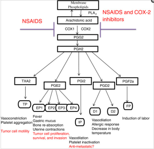
Three important kinds of enzymes that are activated by stress and radiation are phospholipases (that release fatty acids), tryptophan hydroxylase (that controls the conversion of tryptophan to serotonin), and aromatase (estrogen synthetase, that converts androgens to estrogen). The products of these enzymes stimulate cell division, and produce features of the inflammatory process, including the leakiness of capillaries. -Ray Peat, PhD

Picture Source

Serotonin itself is toxic to nerves, and is part of the adaptive system that gets out of control during prolonged inflammation. Serotonin is an important activator of the phospholipases. -Ray Peat, PhD

People who take aspirin, drink coffee, and use tobacco, have a much lower incidence of Alzheimer’s disease than people who don’t use those things. Caffeine inhibits brain phospholipase, making it neuroprotective in a wide spectrum of conditions. In recent tests, aspirin has been found to prevent the misfolding of the prion protein, and even to reverse the misfolded beta sheet conformation, restoring it to the harmless normal conformation. Nicotine might have a similar effect, preventing deposition of amyloid fibrils and disrupting those already formed (Ono, et al., 2002). Vitamin E, aspirin, progesterone, and nicotine also inhibit phospholipase, which contributes to their antiinflammatory action. Each of the amyloid-forming proteins probably has molecules that interfere with its toxic accumulation. -Ray Peat, PhD

J Biochem. 2002 Mar;131(3):285-92.
Phospholipase A2.
Murakami M, Kudo I.
Phospholipase A2 (PLA2) catalyzes the hydrolysis of the sn-2 position of membrane glycerophospholipids to liberate arachidonic acid (AA), a precursor of eicosanoids including prostaglandins (PGs) and leukotrienes (LTs). The same reaction also produces lysophosholipids, which represent another class of lipid mediators. So far, at least 19 enzymes that possess PLA2 activity have been identified in mammals. The secretory PLA2 (sPLA2) family, in which 10 isozymes have been identified, consists of low-molecular-weight, Ca2+-requiring, secretory enzymes that have been implicated in a number of biological processes, such as modification of eicosanoid generation, inflammation, host defense, and atherosclerosis. The cytosolic PLA2 (cPLA2) family consists of 3 enzymes, among which cPLA2alpha plays an essential role in the initiation of AA metabolism. Intracellular activation of cPLA2alpha is tightly regulated by Ca2+ and phosphorylation. The Ca2+-independent PLA2 (iPLA2) family contains 2 enzymes and may play a major role in membrane phospholipid remodeling. The platelet-activating factor (PAF) acetylhydrolase (PAF-AH) family represents a unique group of PLA2 that contains 4 enzymes exhibiting unusual substrate specificity toward PAF and/or oxidized phospholipids. In this review, we will overview current understanding of the properties and functions of each enzyme belonging to the sPLA2, cPLA2, and iPLA2 families, which have been implicated in signal transduction.

Crit Rev Immunol. 1997;17(3-4):225-83.
Regulatory functions of phospholipase A2.
Murakami M, Nakatani Y, Atsumi G, Inoue K, Kudo I.
Phospholipase A2 (PLA2) plays crucial roles in diverse cellular responses, including phospholipid digestion and metabolism, host defense and signal transduction. PLA2 provides precursors for generation of eicosanoids, such as prostaglandins (PGa) and leukotrienes (LTs), when the cleaved fatty acid is arachidonic acid, platelet-activating factor (PAF) when the sn-1 position of the phosphatidylcholine contains an alkyl ether linkage and some bioactive lysophospholipids, such as lysophosphatidic acid (lysoPA). As overproduction of these lipid mediators causes inflammation and tissue disorders, it is extremely important to understand the mechanisms regulating the expression and functions of PLA2. Recent advances in molecular and cellular biology have enabled us to understand the molecular nature, possible function, and regulation of a variety of PLA2 isozymes. Mammalian tissues and cells generally contain more than one enzyme, each of which is regulated independently and exerts distinct functions. Here we classify mammalian PLA2s into there large groups, namely, secretory (sPLA2), cytosolic (cPLA2), and Ca(2+)-independent PLA2s, on the basis of their enzymatic properties and structures and focus on the general understanding of the possible regulatory functions of each PLA2 isozyme. In particular, the roles of type II sPLA2 and cPLA2 in lipid mediator generation are discussed.

C R Seances Soc Biol Fil. 1996;190(4):409-16.
[Diversity of phospholipases A2 and their functions].
[Article in French]
Bereziat G.
Membrane phospholipids not only constitute structural membrane components, they also contain a wealth of biochemical information. They are the source of numerous lipid mediators (prostaglandins, leukotrienes, thromboxane, paf, lysophosphatidic acid and free fatty acids). These lipids act as second messengers inside the cell to modulate enzyme (e.g. PKC and GAP), ion channels (e.g. Ca2+ and K+) or the activity of factors regulating gene expression either at the transcriptional level (e.g. on the TNF alpha gene) or at the post-transcriptional level (e.g. on the GLUT4 transporter). The synthesis of lipid mediators results from the stimulation of phospholipase A2 (PLA2) activities. PLA2 cleaves membrane phospholipids to give rise to lysophospholipids and to free fatty acids from which second messengers are generated. More specifically, PLA2 provides the precursor for the eicosanoids, when the cleaved fatty acid is arachidonic acid, or for PAF, when the sn-1 position of the phospholipid is an alkyl ether linkage. Therefore, PLA2 is a key enzyme in the regulation of lipid mediators of inflammatory process. The purification and cloning of several PLA2s have demonstrated clear differences between secreted and intracellular PLA2. The secreted PLA2s are closely related proteins of low molecular weight (14 kDa) with calcium requirement in the mM range. They contain numerous bonds and retain the same amino-acids at the active site. In mammals, two types of secreted PLA2 have been identified: type I pancreatic PLA2 and type II inflammatory PLA2 which show 70% sequence homology. Recently, two others 14 kDa sPLA2 have been cloned which share also high homologies with type I and type II but contain respectively 6 and 8 disulpide bonds. In contrast, cellular PLA2s have higher molecular weights (40-110 kDa) and are either calcium independent or require microM amounts for activity. Cellular PLA2s preferentially act on sn-2-arachidonoyl phospholipids in vitro whereas sPLA2 do not display such selectivity in vitro. Both cellular and secreted PLA2s are involved in lipid mediator production. Cellular PLA2 can be activated by membrane receptors coupled to G proteins or by tyrosine kinase receptor, through the ras-raf1-MAP kinases network. Cellular PLA2s are thought to be involved in the initial production of lipid mediators after cell activation. Several lines of evidence suggest that secreted PLA2 is involved in the sustained production of lipid mediators in several cell types. These lines of evidence include the decrease in eicosanoid production by antibodies RNA of sPLA2. Furthermore, secreted PLA2s might trigger autocrine loops and proliferation responses through interaction with a specific receptor.

Biochem J. 1994 Aug 1;301 ( Pt 3):721-6.
Fatty acid and phospholipid selectivity of different phospholipase A2 enzymes studied by using a mammalian membrane as substrate.
Diez E, Chilton FH, Stroup G, Mayer RJ, Winkler JD, Fonteh AN.
Previous studies using phospholipid mixed vesicles have demonstrated that several types of phospholipase A2 (PLA2) enzymes exhibit different selectivity for fatty acids at the sn-2 position, for the type of chemical bond at the sn-1 position or for the phosphobase moiety at the sn-3 position of phospholipids. In the present study, we have utilized natural mammalian membranes from U937 monocytes to determine whether two purified 14 kDa PLA2 isoenzymes (Type I, Type II) and a partially purified 110 kDa PLA2 exhibit substrate selectivity for certain fatty acids or phospholipids. In these studies, arachidonic acid (AA) release from membranes was measured under conditions where the remodelling of AA mediated by CoA-independent transacylase (CoA-IT) activity has been eliminated. In agreement with the mixed-vesicle models, AA was the major unsaturated fatty acid hydrolysed from membranes by the 110 kDa PLA2, suggesting that this PLA2 is selective in releasing AA from natural membranes. By contrast, Type I and Type II PLA2s were less selective in releasing AA from phospholipids and released a variety of unsaturated fatty acids at molar ratios that were proportional to the ratios of these fatty acids in U937 microsomal membranes. Examination of AA release from phospholipid classes indicated that all three enzymes released AA from the major AA-containing phospholipid classes (phosphatidylethanolamine, phosphatidylcholine, and phosphatidylinositol) of U937 membranes. The 110 kDa PLA2 released AA from phospholipid subclasses in ratios that were proportional to the AA content within phospholipid classes and subclasses of U937 membranes. These data suggested that the 110 kDa PLA2 shows no preference either for the sn-1 linkage or for the sn-3 phosphobase moiety of phospholipids. By contrast, Type I and Type II PLA2s preferentially released AA from ethanolamine-containing phospholipids and appeared to prefer the 1-acyl-linked subclass. Taken together, these data indicate that the 110 kDa PLA2 selectively releases AA from U937 membranes, whereas Type I and Type II PLA2 release a variety of unsaturated fatty acids. Furthermore, the 110 kDa PLA2 releases the same molar ratios of AA from all major phospholipid subclasses, whereas Type I and Type II PLA2s show some specificity for phosphatidylethanolamine when these enzymes are incubated with a complex mammalian membrane substrate.

J Neurotrauma. 1995 Oct;12(5):791-814.
Mediators of injury in neurotrauma: intracellular signal transduction and gene expression.
Bazan NG, Rodriguez de Turco EB, Allan G.
Membrane lipid-derived second messengers are generated by phospholipase A2 (PLA2) during synaptic activity. Overstimulation of this enzyme during neurotrauma results in the accumulation of bioactive metabolites such as arachidonic acid, oxygenated derivatives of arachidonic acid, and platelet-activating factor (PAF). Several of these bioactive lipids participate in cell damage, cell death, or repair-regenerative neural plasticity. Neurotransmitters may activate PLA2 directly when linked to receptors coupled to G proteins and/or indirectly as calcium influx or mobilization from intracellular stores is stimulated. The release of arachidonic acid and its subsequent metabolism to prostaglandins are early responses linked to neuronal signal transduction. Free arachidonic acid may interact with membrane proteins, i.e., receptors, ion channels, and enzymes, modifying their activity. It can also be acted upon by prostaglandin synthase isoenzymes (the constitutive prostaglandin synthase PGS-1 or the inducible PGS-2) and by lipoxygenases, with the resulting formation of different prostaglandins and leukotrienes. Glutamatergic synaptic activity and activation of postsynaptic NMDA receptors are examples of neuronal activity, linked to memory and learning processes, which activate PLA2 with the consequent release of arachidonic acid and platelet-activating factor (PAF), another lipid mediator. Both mediators may exert presynaptic and postsynaptic effects contributing to long-lasting changes in glutamate synaptic efficacy or long-term potentiation (LTP), PAF, a potential retrograde messenger in LTP, stimulates glutamate release. The PAF antagonist BN 52021 competes for receptors in presynaptic membranes and blocks this effect. PAF may also be involved in plasticity responses because PAF leads to the expression of early response genes and subsequent gene cascades. The PAF antagonist BN 50730, selective for PAF intracellular binding, blocks PAF-mediated induction of gene expression. A consequence of neural injury induced by ischemia, trauma, or seizures is an increased release of neurotransmitters, that in turn generates an overproduction of second messengers. Glutamate, a key player in excitotoxic neuronal damage, triggers increased permeation of calcium mediated by NMDA receptors and activation of PLA2 in postsynaptic neurons. NMDA receptor antagonists reduce the accumulation of free fatty acids and elicit neuroprotection in ischemic damage. Increased production of free arachidonic acid and PAF converges to exacerbate glutamate-mediated neurotransmission. These neurotoxic actions may be brought about by arachidonic acid-induced potentiation of NMDA receptor activity and decreased glutamate reuptake. On the other hand, PAF stimulates the further release of glutamate at presynaptic endings. The neuroprotective effects of the PAF antagonist BN 52021 in ischemia-reperfusion are due, at least in part, to an inhibition of presynaptic glutamate release. PAF also induces expression of the inducible prostaglandin synthase gene, and PAF antagonists selective for the intracellular sites inhibit this effect. The PAF antagonist also inhibits the enhanced abundance, due to vasogenic cerebral edema and ischemia-reperfusion damage, of inducible prostaglandin synthase mRNA in vivo. Therefore, PAF, an injury-generated mediator, may favor the formation of other cell injury and inflammation mediators by turning on the expression of the gene that encodes prostaglandin synthase.

Drug Discov Today. 2003 Aug 1;8(15):710-6.
Phospholipase A2 expression in tumours: a target for therapeutic intervention?
Laye JP, Gill JH.
Phospholipase A(2) (PLA(2)) enzymes are involved in lipid metabolism and, as such, are central to several cellular processes. The different PLA(2)s identified to date can be classified into three groups: secreted PLA(2) (sPLA(2)), calcium-independent PLA(2) (iPLA(2)) and calcium-dependent cytosolic PLA(2) (cPLA(2)). In addition to their role in cellular signalling, PLA(2)s have been implicated in diverse pathological conditions, including inflammation, tissue repair and cancer. Elevated levels of sPLA(2) and cPLA(2) have been reported in several tumour types. Here, we summarize the current views on the PLA(2)s, and look at their expression, role in human malignancy and potential as targets for anticancer drug development.

Screenshot 2017-06-14 at 6.36.36 AM

Picture Source

Curr Mol Med. 2001 Dec;1(6):739-54.
Mammalian secreted phospholipases A2 and their pathophysiological significance in inflammatory diseases.
Touqui L, Alaoui-El-Azher M.
Phospholipases A2 (PLA2s) represent a growing family of enzymes that catalyze the hydrolysis of phospholipids at the sn-2 position leading to the generation of free fatty acids and lysophospholipids. Mammalian PLA2s are divided into two major classes according to their molecular mass and location: intracellular PLA2 and secreted PLA2 (sPLA2). Type-IIA sPLA2 (sPLA2-IIA), the best studied enzyme of sPLA2, plays a role in the pathogenesis of various inflammatory diseases. Conversely, sPLA2-IIA can exert beneficial action in the context of infectious diseases since recent studies have shown that this enzyme exhibits potent bactericidal effects. Induction of the synthesis of sPLA2-IIA is generally initiated by endotoxin and a limited number of cytokines via paracrine and/or autocrine processes. If the mechanisms involved in the regulation of sPLA2-IIA gene expression have been relatively clarified, little is known on the mechanisms that regulate the expression of other sPLA2. There have been substantial progresses in understanding the transcriptional regulation of sPLA2-IIA expression. Recently, transcription factors including NF-kappaB, PPAR, C/EBP have been identified to play a prominent role in the regulation of sPLA2-IIA gene expression. The activation of these transcription factors is under the control of distinct signaling pathways (PKC, cAMP …). Accumulating evidences in the literature suggest that cytosolic PLA2 together with two sPLA2 isozymes (sPLA2-IIA and sPLA2-V) are functionally coupled with cyclooxygenase-1 and 2 pathways, respectively, for immediate and delayed PG biosynthesis. This spatio-temporal coupling of cyclooxygenase enzymes with PLA2s may represent a key mechanism in the propagation of inflammatory reaction. Unraveling the mechanisms involved in the regulation of the expression of sPLA2s is important for understanding their pathophysiological roles in inflammatory diseases.

J Biol Chem. 1995 Jun 23;270(25):14855-8.
Nitric oxide activates the glucose-dependent mobilization of arachidonic acid in a macrophage-like cell line (RAW 264.7) that is largely mediated by calcium-independent phospholipase A2.
Gross RW, Rudolph AE, Wang J, Sommers CD, Wolf MJ.
Herein, we demonstrate that nitric oxide is a potent (> 20% release) and highly selective inducer of [3H]arachidonic acid mobilization in the macrophage-like cell line RAW 264.7. Treatment of RAW 264.7 cells with (E)-6-(bromomethylene)-3-(1-naphthalenyl)-2H-tetrahydropyran-2-one resulted in the inhibition of the large majority (86%) of nitric oxide-induced [3H]arachidonic acid release into the medium (IC50 < 0.5 microM) and the concomitant inhibition of in vitro measurable calcium-independent phospholipase A2 activity (92% inhibition) without demonstrable effects on calcium-dependent phospholipase A2 activity. Since nitric oxide is a potent stimulator of glycolysis (and therefore glycolytically derived ATP) and since cytosolic calcium-independent phospholipase A2 exists as a catalytic complex comprised of ATP-modulated phosphofructokinase-like regulatory polypeptides and a catalytic subunit, we examined the role of glucose in facilitating nitric oxide-mediated arachidonic acid release. Nitric oxide-induced release of [3H]arachidonic acid possessed an obligatory requirement for glucose, was highly correlated with the concentration of glucose in the medium, and was dependent on the metabolism of glucose. Thus, [3H]arachidonic acid release is coupled to cellular glucose metabolism through alterations in the activity of calcium-independent phospholipase A2. Collectively, these results identify a unifying metabolic paradigm in which the generation of lipid second messengers is coordinately linked to the signalstimulated acceleration of glycolytic flux, thereby facilitating integrated metabolic responses to cellular stimuli.

Estrogen, by activating phospholipase A2, acts to amplify the toxic effects of PUFA in the tissues, and these effects increase with age, and with decreased amounts of thyroid and progesterone. -Ray Peat, PhD

Estrogen increases lipid peroxidation, and maintains a chronically high circulating level of free fatty acids, mainly PDFA, activates the phospholipases that release arachidonic acid from cells leading to formation of prostaglandins and isoprostanes, and increases the enzymes that form the inflammation-promoting platelet activating factor (PAF) while suppressing the enzymes that destroy it, and increases a broad range of other inflanunatory mediators, interleukins, and NF-kappa B. -Ray Peat, PhD

Steroids. 2006 Mar;71(3):256-65. Epub 2005 Dec 22.
Estrogen induces phospholipase A2 activation through ERK1/2 to mobilize intracellular calcium in MCF-7 cells.
Thomas W, Coen N, Faherty S, Flatharta CO, Harvey BJ.
The principal secreted estrogen, 17beta-estradiol rapidly activates signaling cascades that regulate important physiological processes including ion transport across membranes, cytosolic pH and cell proliferation. These effects have been extensively studied in the MCF-7 estrogen-responsive human breast carcinoma cell line. Here, we demonstrate that a physiological concentration of 17beta-estradiol caused a rapid, synchronous and transient increase in intracellular calcium concentration in a confluent monolayer of MCF-7 cells 2-3 min after treatment. This response was abolished when cells were pre-incubated with the phospholipase A(2) (PLA(2)) inhibitor quinacrine or with the cyclooxygenase inhibitor indomethacin. The translocation of GFP-cPLA(2)alpha to perinuclear membranes occurred 1-2 min after 17beta-estradiol treatment; this translocation was concurrent with the transient phosphorylation of cPLA(2)alpha at serine residue 505. The phosphorylation and translocation of cPLA(2) were sensitive to inhibition of the extracellular signal regulated kinase (ERK) signaling cascade and occurred simultaneously with a transient activation of ERK. The phosphorylation of cPLA(2) could be stimulated by membrane impermeable 17beta-estradiol conjugated to bovine serum albumen and was blocked by an antagonist of the classical estrogen receptor. Here we show, for the first time, that PLA(2) and the eicosanoid biosynthetic pathway are involved in the 17beta-estradiol induced rapid calcium responses of breast cancer cells.

Prostaglandins. 1996 Mar;51(3):191-201.
Effect of hormones and antihormones on phospholipase A2 activity in human endometrial stromal cells.
Periwal SB, Farooq A, Bhargava VL, Bhatla N, Vij U, Murugesan K.
Phospholipase A2 activity was studied in isolated human endometrial predecidual cells, and in human endometrium collected from day 19-23 of the menstrual cycle, by performing a radiochemical assay. Phospholipase A2 activity on day 20 was significantly higher than other days (P < 0.001), and the activity was found to gradually decrease after day 20 of the menstrual cycle. The effects of the hormones estradiol and progesterone, and antihormones tamoxifen and RU 486, were studied on the phospholipase A2 activity in isolated predecidual stromal cells. Estradiol produced a significant stimulatory effect (P < 0.001) on phospholipase A2 activity in predecidual cells, and this effect was antagonized by tamoxifen. The combination of estradiol and tamoxifen was significantly different from estradiol alone (P < 0.001), but not from tamoxifen alone. RU 486 alone significantly increased (P < 0.001) phospholipase A2 activity in predecidual stromal cells. However, progesterone had no effect on phospholipase A2 activity in predecidual stromal cells.

If the cells adapt to the increased calcium, rather than dying, their sensitivity is reduced. This is probably involved in the “defensive inhibition” seen in many types of cell. In the brain, DHA and arachidonic acid “brought the cells to a new steady state of a moderately elevated [intracellular calcium] level, where the cells became virtually insensitive to external stimuli. This new steady state can be considered as a mechanism of self protection” (Sergeeva, et al., 2005). -Ray Peat, PhD

Reprod Nutr Dev. 2005 Sep-Oct;45(5):633-46.
Regulation of intracellular calcium levels by polyunsaturated fatty acids, arachidonic acid and docosahexaenoic acid, in astrocytes: possible involvement of phospholipase A2.
Sergeeva M, Strokin M, Reiser G.
Pathological conditions in the brain, such as ischemia, trauma and seizure are accompanied by increased levels of free n-6 and n-3 polyunsaturated fatty acids (PUFA), mainly arachidonic acid (AA, 20:4n-6) and docosahexaenoic acid (DHA, 22:6n-3). A neuroprotective role has been suggested for PUFA. For investigation of the potential molecular mechanisms involved in neuroprotection by PUFA, we studied the regulation of the concentration of intracellular Ca2+ ([Ca2+]i) in rat brain astrocytes. We evaluated the presence of extracellular PUFA and the release of intracellular PUFA. Interestingly, only the constitutive brain PUFA AA and DHA, but not eicosapentaenoic acid (EPA) had prominent effects on intracellular Ca2+. AA and DHA suppressed [Ca2+]i oscillation, inhibited store-operated Ca2+ entry, and reduced the amplitudes of Ca2+ responses evoked by agonists of G protein-coupled receptors. Moreover, prolonged exposure of astrocytes to AA and DHA brought the cells to a new steady state of a moderately elevated [Ca2+]i level, where the cells became virtually insensitive to external stimuli. This new steady state can be considered as a mechanism of self-protection. It isolates disturbed parts of the brain, because AA and DHA reduce pathological overstimulation in the tissue surrounding the damaged area. In inflammation-related events, frequently AA and DHA exhibit opposite effects. However, in astrocytes AA and DHA exerted comparable effects on [Ca2+]i. Extracellularly added AA and DHA, but not EPA, were also able to induce the release of [3H]AA from prelabeled astrocytes. Therefore, we also suggest the involvement of phospholipase A2 activation and lysophospholipid generation in the regulation of intracellular Ca2+ in astrocytes.

Increasing intracellular calcium activates phospholipases, releasing more polyunsaturated fats (Sweetman, et al., 1995). -Ray Peat, PhD

Arch Biochem Biophys. 1995 Oct 20;323(1):97-107.
Effect of linoleic acid hydroperoxide on endothelial cell calcium homeostasis and phospholipid hydrolysis.
Sweetman LL, Zhang NY, Peterson H, Gopalakrishna R, Sevanian A.

J Biol Chem 1995 Jun 23;270(25):14855-8.
Nitric oxide activates the glucose-dependent mobilization of arachidonic acid in a macrophage-like cell line (RAW 264.7) that is largely mediated by calcium-independent phospholipase A2.
Gross RW; Rudolph AE; Wang J; Sommers CD; Wolf MJ.
Herein, we demonstrate that nitric oxide is a potent (> 20% release) and highly selective inducer of [3H]arachidonic acid mobilization in the macrophage-like cell line RAW 264.7. Treatment of RAW 264.7 cells with (E)-6-(bromomethylene)-3-(1-naphthalenyl)-2H-tetrahydropyran-2-one resulted in the inhibition of the large majority (86%) of nitric oxide-induced [3H]arachidonic acid release into the medium (IC50 < 0.5 microM) and the concomitant inhibition of in vitro measurable calcium-independent phospholipase A2 activity (92% inhibition) without demonstrable effects on calcium-dependent phospholipase A2 activity. Since nitric oxide is a potent stimulator of glycolysis (and therefore glycolytically derived ATP) and since cytosolic calcium-independent phospholipase A2 exists as a catalytic complex comprised of ATP-modulated phosphofructokinase-like regulatory polypeptides and a catalytic subunit, we examined the role of glucose in facilitating nitric oxide-mediated arachidonic acid release. Nitric oxide-induced release of [3H]arachidonic acid possessed an obligatory requirement for glucose, was highly correlated with the concentration of glucose in the medium, and was dependent on the metabolism of glucose. Thus, [3H]arachidonic acid release is coupled to cellular glucose metabolism through alterations in the activity of calcium-independent phospholipase A2. Collectively, these results identify a unifying metabolic paradigm in which the generation of lipid second messengers is coordinately linked to the signal stimulated acceleration of glycolytic flux, thereby facilitating integrated metabolic responses to cellular stimuli.

Proc Natl Acad Sci U S A 1990 Nov;87(22):8845-9.
Incorporation of marine lipids into mitochondrial membranes increases susceptibility to damage by calcium and reactive oxygen species: evidence for enhanced activation of phospholipase A2 in mitochondria enriched with n-3 fatty Acids.
Malis CD, Weber PC, Leaf A, Bonventre JV.
Experiments were designed to evaluate the susceptibility of mitochondrial membranes enriched with n-3 fatty acids to damage by Ca2+ and reactive oxygen species. Fatty acid content and respiratory function were assessed in renal cortical mitochondria isolated from fish-oil- and beef-tallow-fed rats. Dietary fish oils were readily incorporated into mitochondrial membranes. After exposure to Ca2+ and reactive oxygen species, mitochondria enriched in n-3 fatty acids, and using pyruvate and malate as substrates, had significantly greater changes in state 3 and uncoupled respirations, when compared with mitochondria from rats fed beef tallow. Mitochondrial site 1 (NADH coenzyme Q reductase) activity was reduced to 45 and 85% of control values in fish-oil- and beef-tallow-fed groups, respectively. Exposure to Ca2+ and reactive oxygen species enhance the release of polyunsaturated fatty acids enriched at the sn-2 position of phospholipids from mitochondria of fish-oil-fed rats when compared with similarly treated mitochondria of beef-tallow-fed rats. This release of fatty acids was partially inhibited by dibucaine, the phospholipase A2 inhibitor, which we have previously shown to protect mitochondria against damage associated with Ca2+ and reactive oxygen species. The results indicate that phospholipase A2 is activated in mitochondria exposed to Ca2+ and reactive oxygen species and is responsible, at least in part, for the impairment of respiratory function. Phospholipase A2 activity and mitochondrial damage are enhanced when mitochondrial membranes are enriched with n-3 fatty acids.

Serum amyloid A, which can increase 1000-fold under the influence of proinflammatory cytokines, resulting from irradiation, stress, trauma, or infection, is an activator of phospholipase A2 (PLA2), which releases fatty acids. Some of the neurodegenerative states, including amyloid-prion diseases, involve activated PLA2, as well as increases in the toxic breakdown products of the polyunsaturated fatty acids, such as 4-hydroxynonenal. The quantity of PUFA in the tissues strongly determines the susceptibility of the tissue to injury by radiation and other stresses. But a diet rich in PUFA will produce brain damage even without exceptional stressors, when there aren’t enough antioxidants, such as vitamin E and selenium, in the diet. -Ray Peat, PhD

J Biol Chem. 2004 Aug 27;279(35):36405-11. Epub 2004 Jun 21.
Phospholipase A2 inhibitors or platelet-activating factor antagonists prevent prion replication.
Bate C, Reid S, Williams A.
A key feature of prion diseases is the conversion of the cellular prion protein (PrP(C)) into disease-related isoforms (PrP(Sc)), the deposition of which is thought to lead to neurodegeneration. In this study a pharmacological approach was used to determine the metabolic pathways involved in the formation of protease-resistant PrP (PrP(res)) in three prion-infected cell lines (ScN2a, SMB, and ScGT1 cells). Daily treatment of these cells with phospholipase A(2) (PLA(2)) inhibitors for 7 days prevented the accumulation of PrP(res). Glucocorticoids with anti-PLA(2) activity also prevented the formation of PrP(res) and reduced the infectivity of SMB cells. Treatment with platelet-activating factor (PAF) antagonists also reduced the PrP(res) content of cells, while the addition of PAF reversed the inhibitory effect of PLA(2) inhibitors on PrP(res) formation. ScGT1 cells treated with PLA(2) inhibitors or PAF antagonists for 7 days remained clear of detectable (PrPres) when grown in control medium for a further 12 weeks. Treatment of non-infected cells with PLA(2) inhibitors or PAF antagonists reduced PrP(C) levels suggesting that limiting cellular PrP(C) may restrict prion formation in infected cells. These data indicate a pivotal role for PLA(2) and PAF in controlling PrP(res) formation and identify them as potential therapeutic agents.

Zhongguo Yao Li Xue Bao. 1994 Jul;15(4):299-302.
Three drugs inhibit phospholipase A2-induced high permeability of endothelial monolayers.
Chen SF, Li SH, Ding FY.
The permeability of aortic endothelial monolayers to fluid and albumin increased 13.5 and 16.1 times respectively after pretreatment with phospholipase A2 (PLA2, 100 U.ml-1) for 30 min. 1-(p-Chlorobenzoyl)-5-methylindole-3-acetic acid (1.16 mmol.L-1), SRI 63-441 (30 nmol.L-1), and 1,25-dihydroxycholecalciferol (0.1 mumol.L-1) decreased PLA2-induced high permeability. PLA2 did not damage the endothelial cells significantly. Our results indicate that the action of PLA2 to increase the permeability of endothelial monolayers is mainly due to PLA2-induced lipid mediators released from endothelial cells.

Inflammation. 1990 Jun;14(3):267-73.
Phospholipase A2-induced lung edema and its mechanism in isolated perfused guinea pig lung.
Chen SF, Li SH, Fei X, Wu ZL.
Lung injury induced by phospholipase A2 (PLA2, 0.046 IU/ml perfusate) was studied in a continuous weighing system of isolated perfused guinea pig lungs. The results revealed that lung weight increased progressively during the 30-min perfusion of PLA2. No change of pulmonary arterial pressure was observed in the same period. Albumin permeability-surface area product, lung index, lung water content, exudate from pleura, and angiotensin-converting-enzyme activity increased significantly at the end of 30 min PLA2 perfusion. p-Bromophenacyl bromide, a PLA2 inhibitor, may block the above changes nearly completely. The effects of inhibitors of cyclooxygenase (indomethacin, IM), lipoxygenase (diethylcarbamaxine, DE), and platelet-activating factor (SRI 63-441) on PLA2-induced lung injury were also studied. We found: (1) PLA2 may induce high permeability lung edema. The role of endothelial injury in the permeability change remains to be further investigated. (2) DE ameliorated lung injury significantly within 10 min of PLA2 treatment but showed no effect after 15 min. IM ameliorated lung injury during the whole experimental period. SRI 63-441 had no effect. It is suggested that PLA2 may damage lung by inducing products of cyclooxygenase and lipoxygenase besides its direct effect.

Am Rev Respir Dis. 1990 Nov;142(5):1193-9.
Phospholipase A2-induced pulmonary and hemodynamic responses in the guinea pig. Effects of enzyme inhibitors and mediators antagonists.
Tocker JE, Durham SK, Welton AF, Selig WM.
The effect of phospholipase A2 (Naja naja) PLA2) on mean arterial blood pressure and intratracheal pressure was examined in anesthetized guinea pigs. Intracheally administered PLA2 (1 to 10 U) produced acute, dose-dependent increases in mean arterial blood pressure and intracheal pressure. However, Intravenously administered PLA2 (doses as large as 1,000 U) did not alter monitored variables. Acute PLA2-induced morphologic alterations were characterized by airway constriction, airway/alveolar cell damage, and pulmonary sequestration of both leukocytes and platelets. PLA2-induced increases in both mean arterial blood pressure and intratracheal pressure were attenuated to varying degrees by pretreating intravenously with indomethacin (10 mg/kg), a cyclooxygenase inhibitor, and WEB 2086 (0.1 mg/kg), a platelet-activating factor antagonist. Both ICI 198,615 (1 mg/kg), a leukotriene D4, receptor antagonist given intravenously, and dexamethasone (50 mg/kg), a steroidal anti-inflammatory agent given intraperitoneally as a 2-day pretreatment, reduced PLA2-induced increases in intratracheal pressure. Pyrilamine (2 mg/kg), a histamine1-receptor antagonist given intravenously, did not modify PLA2-induced pathophysiologic responses. Guinea pigs exposed to aerosolized PLA2 (100 U/ml) exhibited evidence of increased bronchoalveolar lavage macrophage, leukocyte, and lymphocyte accumulation at 24 h post-PLA2. These studies suggest that in vivo PLA2-induced pathophysiologic changes in the guinea pig involve alterations in resident airway cell populations as well as sequestration and infiltration of inflammatory cells. Both eicosanoids and platelet-activating factor appear to contribute to these PLA2-induced pathophysiologic effects.

Prostaglandins Leukot Essent Fatty Acids. 1990 Mar;39(3):167-75.
The effects of neutrophils and phospholipase A2 on transvascular albumin flux in isolated rabbit lungs.
Littner MR, Lott FD.
In this study, addition of phospholipase A2 (PLA2) to salt-perfused isolated rabbit lungs containing rabbit polymorphonuclear leukocytes leads to an increase in pulmonary capillary permeability. We add 1.5 X 10(8) polymorphonuclear leukocytes to the perfusate. Next, indomethacin is added to the perfusate and 40 units of PLA2 are infused into the pulmonary arterial inflow of the lungs. At the end of the study, a lung sample is removed for measurement of transvascular albumin flux using I125-albumin as a measure of the permeability-surface area product. Control studies demonstrate no increase in transvascular albumin flux. Addition of a dual cyclooxygenase and lipoxygenase inhibitor, BW755C, to the perfusate prevents the increase in transvascular albumin flux. We conclude that PLA2 interacts with polymorphonuclear leukocytes to increase protein permeability. Since PLA2 can release endogenous arachidonic acid and platelet-activating factor from cells, this suggests that release of such products may contribute to an increase in pulmonary capillary permeability from polymorphonuclear leukocytes. The ability of BW755C to prevent the increase suggests the possibility that lipoxygenase products contribute.

Am J Pathol. 1990 Jun;136(6):1283-91.
Phospholipase A2-induced pathophysiologic changes in the guinea pig lung.
Durham SK, Selig WM.
The pathophysiology of lung injury induced by phospholipase A2 (PLA2), a lipolytic enzyme implicated in a variety of pulmonary diseases, was examined in the guinea pig. One hundred microliters of saline or 10 units of PLA2 suspended in saline was given as a bolus injection into either the trachea or jugular vein. Intratracheal pressure and mean arterial blood pressure were continuously monitored. The lungs were examined by light and transmission electron microscopy at 1, 10, and 30 minutes after administration. Pulmonary morphologic and physiologic changes were only observed in animals that received PLA2 via the trachea. Significant increases in peak intratracheal pressure occurred as early as 1 minute after intratracheal PLA2 administration. Morphologic evidence of airway constriction, accompanied by blebbing of the apical cytoplasm of airway epithelium, was also observed at this time. A transient increase in mean arterial blood pressure occurred 5 minutes after challenge. At 10 minutes after intratracheal PLA2, there was marked swelling of airway epithelial cells, pronounced blebbing of the apical cytoplasm, and a resultant decrease in size of the airway lumen. Morphologic changes in alveolar cell populations were initially observed 10 minutes after intratracheal PLA2. Interalveolar septa were hypercellular and multifocally thickened. There was prominent perivascular edema and alveolar spaces contained abundant proteinaceous material and occasional hemorrhage. Ultrastructurally, there was marked cell swelling and fragmentation of type I alveolar epithelium resulting in a denuded basal lamina. Sequestration of neutrophils and eosinophils, many of which lacked secretory granules, within alveolar capillaries was accompanied by aggregates of platelets and was observed in close proximity to injured endothelium. Morphologic changes indicative of cell injury were also observed in type II alveolar epithelium. Similar, but more frequent and severe, morphologic injury occurred 30 minutes after intratracheal PLA2. It is concluded that PLA2 induces pronounced morphologic and physiologic changes in the guinea pig and that the route of administration is important in the development of PLA2-induced lung injury.

Thorax. 2008 Jan;63(1):21-6. Epub 2007 Jun 15.
Plasma phospholipase A2 activity in patients with asthma: association with body mass index and cholesterol concentration.
Misso NL, Petrovic N, Grove C, Celenza A, Brooks-Wildhaber J, Thompson PJ.
BACKGROUND:
Secretory phospholipases A2 (sPLA2) have functions relevant to asthmatic inflammation, including eicosanoid synthesis and effects on dendritic cells and T cells. The aim of this study was to measure sPLA2 activity in patients with stable and acute asthma and to assess potential associations with body mass index (BMI), and plasma cholesterol and vitamin C concentrations.
METHODS:
Plasma sPLA2 activity and concentrations of cholesterol and vitamin C were measured in 23 control subjects and 61 subjects with stable asthma (42 mild to moderate, 19 severe). In addition, sPLA2 activity was measured in 36 patients experiencing acute asthma and in 22 of these patients after recovery from the acute attack.
RESULTS:
sPLA2 activity was not significantly greater in severe (499.9 U; 95% confidence interval (CI) 439.4 to 560.4) compared with mild to moderate asthmatic subjects (464.8; 95% CI 425.3 to 504.3) or control subjects (445.7; 95% CI 392.1 to 499.4), although it was higher in patients with acute asthma (581.6; 95% CI 541.2 to 622.0; p<0.001). Male gender, high plasma cholesterol, increased BMI and atopy were associated with increased sPLA2 activity, while plasma vitamin C was inversely correlated with sPLA2 activity in patients with stable asthma and in control subjects. There were significant interactions between gender and plasma cholesterol and between gender and vitamin C in relation to sPLA2 activity.
CONCLUSIONS:
Plasma sPLA2 may provide a biological link between asthma, inflammation, increased BMI, lipid metabolism and antioxidants. Interactions among these factors may be pertinent to the pathophysiology and increasing prevalence of both asthma and obesity.

Cell Mol Biol (Noisy-le-grand). 2004 Feb;50(1):87-94.
The role of secretory phospholipase A2 in acute chest syndrome.
Kuypers FA, Styles LA.
Acute chest syndrome (ACS) is the leading cause of death in sickle cell disease. Severe ACS often develops in the course of a vasoocclusive crisis (VOC), and frequently involves pulmonary fat embolism. Secretory phospholipase A2 (sPLA2), a potent inflammatory mediator, is elevated in ACS, and sPLA2 levels in serum or plasma predict impending ACS. In addition sPLA2 may play a major role in the actual damage to the lung resulting in a new pulmonary infiltrate on chest radiography, respiratory symptoms, and ultimately alveolar collapse and the impairment of gas exchange. The data indicate that measurement of sPLA2 can be useful in alerting the clinician to patients with impending ACS, and suggest that instituting early therapies based on sPLA2 levels, including inhibition of sPLA2 activity, may be useful to prevent or reduce the clinical morbidity of ACS in sickle cell disease.

Blood. 1996 Mar 15;87(6):2573-8.
Phospholipase A2 levels in acute chest syndrome of sickle cell disease.
Styles LA, Schalkwijk CG, Aarsman AJ, Vichinsky EP, Lubin BH, Kuypers FA.
Acute chest syndrome (ACS) is associated with significant morbidity and is the leading cause of death in patients with sickle cell disease (SCD). Recent reports suggest that bone marrow fat embolism can be detected in many cases of severe ACS. Secretory phospholipase A2 (sPLA2) is an important inflammatory mediator and liberates free fatty acids, which are felt to be responsible for the acute lung injury of the fat embolism syndrome. We measured SPLA2 levels in 35 SCD patients during 20 admissions for ACS, 10 admissions for vaso-occlusive crisis, and during 12 clinic visits when patients were at the steady state. Eleven non-SCD patients with pneumonia were also evaluated. To determine if there was a relationship between sPLA2 and the severity of ACS we correlated SPLA2 levels with the clinical course of the patient. In comparison with normal controls (mean = 3.1 +/- 1.1 ng/mL), the non-SCD patients with pneumonia (mean = 68.6 +/- 82.9 ng/mL) and all three SCD patient groups had an elevation of SPLA2 (steady state mean = 10.0 +/- 8.4 ng/mL; vaso-occlusive crisis mean = 23.7 +/- 40.5 ng/mL; ACS mean = 336 +/- 209 ng/mL). In patients with ACS sPLA2 levels were 100-fold greater than normal control values, 35 times greater than values in SCD patients at baseline, and five times greater than non-SCD patients with pneumonia. The degree of SPLA2 elevation in ACS correlated with three different measures of clinical severity and, in patients followed sequentially, the rise in SPLA2 coincided with the onset of ACS. The dramatic elevation of SPLA2 in patients with ACS but not in patients with vaso-occlusive crisis or non-SCD patients with pneumonia and the correlation between levels of SPLA2 and clinical severity suggest a role for SPLA2 in the diagnosis and, perhaps, in the pathophysiology of patients with ACS.

Blood. 2000 Nov 1;96(9):3276-8.
Secretory phospholipase A(2) predicts impending acute chest syndrome in sickle cell disease.
Styles LA, Aarsman AJ, Vichinsky EP, Kuypers FA.
Acute chest syndrome (ACS) is the leading cause of death in sickle cell disease. Severe ACS often develops in the course of a vaso-occlusive crisis (VOC), but currently there are no predictors for its development. Secretory phospholipase A(2) (sPLA(2)), a potent inflammatory mediator, is elevated in ACS, and previous work suggests that sPLA(2) predicts impending ACS. We prospectively evaluated sPLA(2) concentration during 21 admissions for VOC; 6 of these patients went on to develop ACS. Elevation of sPLA(2) was detected all 6 patients 24 to 48 hours before ACS was clinically diagnosed. Adding the requirement for fever raised the specificity of sPLA(2) to 87% while retaining 100% sensitivity. These data indicate that sPLA(2) can be useful in alerting the clinician to patients with impending ACS. In addition, sPLA(2) may be useful for instituting early therapies to prevent or reduce the clinical morbidity of ACS.

Am J Respir Crit Care Med. 1997 Feb;155(2):421-5.
Phospholipase A2 and arachidonate increase in bronchoalveolar lavage fluid after inhaled antigen challenge in asthmatics.
Bowton DL, Seeds MC, Fasano MB, Goldsmith B, Bass DA.
Phospholipases A2 (PLA2) hydrolyze phospholipids resulting in the release of fatty acids including arachidonic acid (AA) and lysophospholipids. AA, in turn, serves as a substrate for the synthesis of leukotrienes which can cause bronchoconstriction and airways edema and appear to be important mediators of clinical asthma. Further, lysophospholipids may be cytotoxic and/or impair the function of surfactant. We examined the release of secretory PLA2 (sPLA2) and AA into the airways after antigen challenge in 16 subjects with allergic asthma. Asthmatic subjects underwent bronchoscopy with bronchoalveolar lavage (BAL) before and after inhaled antigen challenge; in addition, a single BAL, without inhaled antigen, was performed in 10 control subjects. BAL was obtained at 4 h (n = 7), the time of the late asthmatic response (LAR) (n = 5), or 24 h (n = 4) after challenge. There was no difference between normal and asthmatic subjects in either BAL fluid (BALF) sPLA2 activity or AA concentration at baseline. Both sPLA2 and AA increased after antigen challenge (p < 0.01 and 0.05, respectively). These changes were most marked 4 h after challenge (p < 0.03 for both). sPLA2 may play an important role in the generation of AA in patients with asthma.

Allergy. 2000;55 Suppl 61:27-30.
Arachidonic acid metabolism in inflammatory cells of patients with bronchial asthma.
Calabrese C, Triggiani M, Marone G, Mazzarella G.
Over the last few years, the demonstration of beneficial effects of leukotriene receptor antagonists in various forms of asthma has renewed clinical and pharmacologic interest in this class of lipid mediators. Several studies demonstrated an increased biosynthesis of cysteinyl leukotrienes (CysLT) in asthmatic patients. However, the reasons for the dysregulated production of CysLTs in asthmatic patients are not completely defined. An improved method of lipid mediator detection and the availability of cells isolated from human airways (by bronchoalveolar lavage [BAL] and bronchial biopsies) have allowed initial studies to address this issue. Eosinophils retrieved from inflamed airways of asthmatics have a larger arachidonic acid (AA) content than their blood counterpart. The high level of AA in these cells is primarily due to a remodeling of endogenous arachidonate pools with the accumulation of this fatty acid in a triglyceride-associated pool. In addition, elevated levels of a secretory form of phospholipase A2, the key enzyme initiating the cascade of CysLTs, are found in the BAL of asthmatics. Finally, eosinophils isolated from the BAL of asthmatics have an increased expression of LTC4 synthase. The level of expression of this enzyme correlates with the increased amount of CysLTs produced in the airways of these patients. Taken together, these data identify at least two possible mechanisms to explain the excessive CysLT production in asthmatics: 1) an increased content of AA in the glycerolipid pools of inflammatory cells 2) an enhanced activity of key biosynthetic enzymes involved in CysLT synthesis.

J Leukoc Biol. 1996 Dec;60(6):704-9.
Secretory phospholipase A2 activity is elevated in bronchoalveolar lavage fluid after ovalbumin sensitization of guinea pigs.
Sane AC, Mendenhall T, Bass DA.
Arachidonic acid (AA), the precursor of eicosanoids, is released from the sn-2 position of phospholipids by both secretory (sPLA2) and cytosolic phospholipase A2 (cPLA2). Eicosanoids have been shown to contribute to bronchospasm in asthma. We measured the enzymatic activity of sPLA2 and cPLA2 in the bronchoalveolar lavage fluid and cells, respectively, in male Hartley guinea pigs sensitized with ovalbumin. sPLA2 activity was also measured from alveolar macrophages (AM) in culture from unsensitized and sensitized animals. There was an increase in sPLA2 activity and AA content in the lavage fluid following sensitization (18.73 +/- 1.33 to 25.74 +/- 3.22% hydrolysis and 17.97 +/- 12.39 to 44.76 +/- 13.37 pmol AA/mL BAL, mean +/- SD), which remained elevated but without further increase 4 or 24 h after antigen challenge. AM from unsensitized and sensitized-unchallenged animals did not secrete sPLA2 activity in culture for 3 h and therefore do not appear to be the cell source of the sPLA2 activity present in the alveolar lavage fluid following OA sensitization. In contrast to the increase in sPLA2 in lung lavage fluid, Western blotting for cPLA2 from lung lavage cells showed no increase 4 or 24 h after antigen challenge compared with sensitization alone. cPLA2 enzymatic activity of the cytosol fraction of lung lavage cells showed no changes with antigen sensitization or challenge. In summary, intraperitoneal sensitization with ovalbumin in male Hartley guinea pigs caused an increase in both sPLA2 and AA in bronchoalveolar lavage fluid without a need for antigen challenge. The increased sPLA2 enzymatic activity following sensitization may be responsible for the elevation of AA in the bronchoalveolar lavage fluid observed after antigen sensitization.

J Thromb Thrombolysis. 2010 Apr;29(3):276-81. Epub 2009 May 17.
Secretory phospholipase A2 in patients with coronary artery disease.
Lima LM, Carvalho MG, da Fonseca Neto CP, Garcia JC, Sousa MO.
This study investigated the correlation of sPLA2 (secretory phospholipase A2) activity with the atheromatosis extent in subjects with coronary artery disease (CAD) undergoing coronary angiography. We analyzed 123 patients, including 35 subjects with angiographically normal coronary arteries (controls), 31 with mild/moderate atheromatosis (stenosis of 30-70% of the luminal diameter in one or more coronary arteries) and 57 with severe atheromatosis (>70% stenosis). Plasma sPLA2 activity was significantly higher in subjects with severe [127.7 U/ml (102.3-162.7); p < 0.0001] and mild/moderate [112.0 U/ml (100.6-146.9); p < 0.0001] atheromatosis than in controls [19.8 U/ml (15.1-32.1)]. In a multiple logistic regression model, adjusted for age, gender, body mass index, tabagism, hypertension, sedentarism, family history for coronary artery disease, diabetes mellitus, total cholesterol, HDLc, LDLc, triglycerides, high sensitivity C-reactive protein and phospholipase A2, only sPLA2 was observed to be independently associated with severe CAD (>70% of stenosis) (p < 0.0001).

Arterioscler Thromb Vasc Biol. 2005 Apr;25(4):839-46. Epub 2005 Feb 3.
Serum levels of type II secretory phospholipase A2 and the risk of future coronary artery disease in apparently healthy men and women: the EPIC-Norfolk Prospective Population Study.
Boekholdt SM, Keller TT, Wareham NJ, Luben R, Bingham SA, Day NE, Sandhu MS, Jukema JW, Kastelein JJ, Hack CE, Khaw KT.
OBJECTIVES:
To study the prospective relationship between serum levels of type II secretory phospholipase A2 (sPLA2) and the risk of future coronary artery disease (CAD) in apparently healthy men and women.
METHODS AND RESULTS:
We conducted a prospective nested case-control study among apparently healthy men and women aged 45 to 79 years. Cases (n=1105) were people in whom fatal or nonfatal CAD developed during follow-up. Controls (n=2209) were matched by age, sex, and enrollment time. sPLA2 levels were significantly higher in cases than controls (9.5 ng/mL; interquartile range [IQR], 6.4 to 14.8 versus 8.3 ng/mL; IQR, 5.8 to 12.6; P<0.0001). sPLA2 plasma levels significantly correlated with age, body mass index, systolic blood pressure, high-density lipoprotein (HDL) cholesterol levels, and C-reactive protein (CRP) levels. Taking into account matching for sex and age and adjusting for body mass index, smoking, diabetes, systolic blood pressure, low-density lipoprotein cholesterol, HDL cholesterol, and CRP levels, the risk of future CAD was 1.34 (1.02 to 1.71; P=0.02) for people in the highest sPLA2 quartile, compared with those in the lowest (P for linearity=0.03).
CONCLUSIONS:
Elevated levels of sPLA2 were associated with an increased risk of future CAD in apparently healthy individuals. The magnitude of the association was similar to that observed between CRP and CAD risk, and both associations were independent.

Increased intracellular calcium activates lipolysis (by phospholipases), producing more free fatty acids, as well as excitation and protein breakdown, and in the brain neurodegenerative diseases, calcium excess contributes to clumping synuclein (Wojda, et al., 2008), an important regulator of cytoskeleton proteins. -Ray Peat, PhD

IUBMB Life. 2008 Sep;60(9):575-90.
Calcium ions in neuronal degeneration.
Wojda U, Salinska E, Kuznicki J.
Neuronal Ca(2+) homeostasis and Ca(2+) signaling regulate multiple neuronal functions, including synaptic transmission, plasticity, and cell survival. Therefore disturbances in Ca(2+) homeostasis can affect the well-being of the neuron in different ways and to various degrees. Ca(2+) homeostasis undergoes subtle dysregulation in the physiological ageing. Products of energy metabolism accumulating with age together with oxidative stress gradually impair Ca(2+) homeostasis, making neurons more vulnerable to additional stress which, in turn, can lead to neuronal degeneration. Neurodegenerative diseases related to aging, such as Alzheimer’s disease, Parkinson’s disease, or Huntington’s disease, develop slowly and are characterized by the positive feedback between Ca(2+) dyshomeostasis and the aggregation of disease-related proteins such as amyloid beta, alfa-synuclein, or huntingtin. Ca(2+) dyshomeostasis escalates with time eventually leading to neuronal loss. Ca(2+) dyshomeostasis in these chronic pathologies comprises mitochondrial and endoplasmic reticulum dysfunction, Ca(2+) buffering impairment, glutamate excitotoxicity and alterations in Ca(2+) entry routes into neurons. Similar changes have been described in a group of multifactorial diseases not related to ageing, such as epilepsy, schizophrenia, amyotrophic lateral sclerosis, or glaucoma. Dysregulation of Ca(2+) homeostasis caused by HIV infection or by sudden accidents, such as brain stroke or traumatic brain injury, leads to rapid neuronal death. The differences between the distinct types of Ca(2+) dyshomeostasis underlying neuronal degeneration in various types of pathologies are not clear. Questions that should be addressed concern the sequence of pathogenic events in an affected neuron and the pattern of progressive degeneration in the brain itself. Moreover, elucidation of the selective vulnerability of various types of neurons affected in the diseases described here will require identification of differences in the types of Ca(2+) homeostasis and signaling among these neurons. This information will be required for improved targeting of Ca(2+) homeostasis and signaling components in future therapeutic strategies, since no effective treatment is currently available to prevent neuronal degeneration in any of the pathologies described here.

J Invest Dermatol. 1993 Jan;100(1):35S-41S.
Mechanisms of UV-induced inflammation.
Hruza LL, Pentland AP.
The inflammation produced by exposure to ultraviolet (UV) light has been well documented clinically and histologically. However, the mechanisms by which mediators induce this clinical response remain poorly defined. It is clear that photochemistry occurring after UV absorption must be responsible for initiating these events. Some of these underlying mechanisms have been defined. After exposure to UV light, the formation of prostaglandins and the release of histamine are increased. In addition to an increase in the quantity of these mediators, an increase in sensitivity of irradiated tissue to agonist stimulation also occurs. This increased sensitivity may cause tissue to respond to agonist levels previously present. Phospholipase activity also increases, making more substrate available for prostaglandin formation. Oxygen radical-induced peroxidation of membrane lipids caused by irradiation may contribute to increased phospholipase activity. Oxygen-free radicals also participate in sunburn cell formation and in UV-induced decreases in Langerhans cell numbers. Several enzymatic and non-enzymatic mechanisms are present in skin for reducing these highly reactive oxygen species.

Posted in General.

Tagged with , , , , , , , , , , , , , , , , , , , , , , , , , , , , , , .


Dietary PUFA Reflected in Human Subcutaneous Fat Tissue

Also see:
Toxicity of Stored PUFA
Vitamin E Needs Increases with PUFA Consumption and Greater Unsaturation
Israeli Paradox: High Omega -6 Diet Promotes Disease
Protective “Essential Fatty Acid Deficiency”
PUFA Accumulation & Aging
Arachidonic Acid’s Role in Stress and Shock
Saturated and Monousaturated Fatty Acids Selectively Retained by Fat Cells
PUFA Promote Stress Response; Saturated Fats Suppress Stress Response
Your MUFA + PUFA Intakes Determine Your True Vitamin E Requirements – N-3s are the Worst Offenders + Even MUFAs Need Buffering | Tool to Calculate Your Individual Needs
The Dangers of Fat Metabolism and PUFA: Why You Don’t Want to be a Fat Burner

Some studies accumulated orginially by Stephan Guyenet, PhD.

Am J Clin Nutr. 1975 Jun;28(6):577-83.
Recommended dietary allowance for vitamin E: relation to dietary, erythrocyte and adipose tissue linoleate.
Witting LA, Lee L.
The general trend toward increased consumption of polyunsaturated fatty acids is apparent in the linoleate level of adipose tissue (13.0 plus or minus 1.3%) and erythrocyte lipids (14.0 plus or minus 1.9%) in the present group of female undergraduate student volunteers compared to values reported in the early 1960’s. On the basis of the level of linoleate in their diets (19.5 plus or minus 0.8%), it is also apparent that further increases in tissue lipid linoleate levels are to be anticipated, which in turn will result in an increased requirement for vitamin E. It is suggested that adipose tissue linoleate levels in the general population be used as a baseline for the periodic evaluation and revision of the recommended dietary allowance for vitamin E. The recommended dietary allowance could then be phrased in terms of the quantity of vitamin E activity to be consumed per gram linoleate in 100 g adipose tissue fatty acids. A recommendation of 0.6 IU vitamin E activity/g linoleate in 100 g adipose tissue fatty acids is tentatively suggested.

Am J Clin Nutr. 1991 Aug;54(2):340-5.
Fatty acid composition of subcutaneous adipose tissue and diet in postmenopausal US women.
London SJ, Sacks FM, Caesar J, Stampfer MJ, Siguel E, Willett WC.
The distributions of fatty acids in subcutaneous-adipose-tissue aspirates and their relation to intake as assessed by a semiquantitative food-frequency questionnaire were investigated in 115 postmenopausal US women free from cancer. Percentages of fatty acids in adipose tissue were significantly correlated with the percentage of total fat intake for polyunsaturated fatty acids (Spearman correlation = 0.37), n-3 fatty acids of marine origin (Spearman correlation = 0.48), and trans fatty acids (Spearman correlation = 0.51) but not for saturated and monounsaturated fatty acids. Correlations were somewhat stronger in 78 women with stable weight over the 6 mo before fat aspiration. These data suggest that intakes of polyunsaturated fatty acids, including n-3 fatty acids, and trans fatty acids are reflected in the adipose tissue but that intakes of saturated and monounsaturated fatty acids are not. The data also provide further support for the validity of the food-frequency questionnaire in the assessment of fat intake.

Am J Clin Nutr. 1998 Jan;67(1):25-30.
The relation between dietary intake and adipose tissue composition of selected fatty acids in US women.
Garland M, Sacks FM, Colditz GA, Rimm EB, Sampson LA, Willett WC, Hunter DJ.
We compared fatty acid amounts in adipose tissue with fatty acid intake calculated from 2 separate weeks of diet recording and two food-frequency questionnaires for 140 participants in the Nurses’ Health Study. Our results showed that the amounts of polyunsaturated and trans fatty acids in adipose tissue reflect dietary intake and confirm those of previous studies. The correlation between the polyunsaturated fatty acid content of adipose tissue and polyunsaturated fatty acid intake from the average of the two food-frequency questionnaires was 0.40; this correlation for trans fatty acids was also 0.40. Linolenic acid amounts in adipose tissue were also moderately correlated with intake from the average of the food-frequency questionnaires (r = 0.34). An estimate of trans fatty acid intake from vegetable sources correlated much more strongly with adipose trans fatty acids than did an estimate of trans fatty acids from animal sources. Adipose tissue aspirates can be used to indicate intake of exogenous fatty acids.

Am J Epidemiol. 1992 Feb 15;135(4):418-27.
Comparison of measures of fatty acid intake by subcutaneous fat aspirate, food frequency questionnaire, and diet records in a free-living population of US men.
Hunter DJ, Rimm EB, Sacks FM, Stampfer MJ, Colditz GA, Litin LB, Willett WC.
In 1986-1987, the authors assessed the fatty acid intake of 118 Boston-area men, aged 40-75 years, by a semiquantitative food frequency questionnaire administered twice, by two 7-day diet records, and by capillary gas chromatography of subcutaneous fat samples obtained by needle aspirate from the lateral buttock. Spearman correlation coefficients between diet record estimates of fatty acid intake (as a percentage of total fat) and fat aspirate measures (as a percentage of total peak area) were as follows: saturated fat, r = 0.16 (p = 0.09); monounsaturated fat, r = 0.22 (p = 0.01); and polyunsaturated fat, r = 0.49 (p = 0.0001). Spearman correlation coefficients between estimates derived from the food frequency questionnaire were as follows: saturated fat, r = 0.18 ( p = 0.05); monounsaturated fat, r = 0.14 (p = 0.14); polyunsaturated fat, r = 0.50 (p = 0.0001); and eicosapentaenoic acid, r = 0.47 (p = 0.0001). These data confirm that the polyunsaturated and eicosapentaenoic fatty acid content of subcutaneous fat is a measure of dietary intake of these fats. Although diet records are commonly thought to be the “gold standard” method of dietary assessment, the similar correlations observed between the fatty acid composition of adipose tissue and estimates of intake from the food frequency questionnaire and from diet records suggest that these two dietary assessment methods have similar validity in the measurement of polyunsaturated fatty acid intake.

Ann Epidemiol. 2003 Feb;13(2):119-27.
Comparison of adipose tissue fatty acids with dietary fatty acids as measured by 24-hour recall and food frequency questionnaire in Black and White Adventists: the Adventist Health Study.
Knutsen SF, Fraser GE, Beeson WL, Lindsted KD, Shavlik DJ.
PURPOSE:
To calibrate and compare intake of different fats and individual fatty acids as assessed with a food frequency questionnaire (FFQ) against that estimated with (i) a series of dietary recalls and; (ii) the relative fat concentration in an adipose tissue biopsy. The FFQ was specially designed for use in a cohort of Seventh-day Adventists. In preparation for a large cohort study investigating the effect of diet on risk of colon, prostate and breast cancer.
METHODS:
The association of adipose tissue fatty acids and dietary fat intake was assessed in 49 black and 72 white Seventh-day Adventists subjects using 8 different 24-hour recalls, a 200-item food frequency questionnaire (FFQ) and adipose tissue biopsies from each subject.
RESULTS:
Pearson correlation between fatty acids in adipose tissue and dietary intake as assessed by multiple 24-hour recalls were as follows: Linoleic acid: 0.77 in black and 0.71 in white subjects, respectively; Linolenic acid: 0.68 (blacks) and 0.62 (whites); Total Polyunsaturated fat (PUFA): 0.78 (blacks) and 0.70 (whites); Total Monounsaturated fat (MUFA): 0.35 (blacks) and 0.03 (whites); Total Saturated fat (SFA): 0.46 (blacks) and 0.56 (whites). Correlations between fatty acids in adipose tissue and dietary intake as assessed by FFQ were: Linoleic acid: 0.61 (blacks) and 0.52 (whites), respectively; Linolenic acid: 0.29 (blacks) and 0.49 (whites); PUFA: 0.62 (blacks) and 0.53 (whites); MUFA: 0.07 (blacks) and 0.31 (whites), SFA: 0.21 (blacks) and 0.31 (whites).
CONCLUSIONS:
Our study confirms findings of others that 24-hour recalls are valid for assessing dietary intake of different types of fat. The FFQ we developed and used in this study gave reasonably valid measures of fatty acid intake in our population and is thus suitable for use in large cohort studies. It had validity comparable to that observed for other FFQs.

Am J Clin Nutr. 1995 Nov;62(5):956-9.
Biomarkers of habitual fish intake in adipose tissue.
Marckmann P, Lassen A, Haraldsdóttir J, Sandström B.
The association between habitual fish and marine n-3 polyunsaturated fatty acid (PUFA) intake, and the fatty acid composition of subcutaneous fat was studied in 24 healthy young volunteers. Habitual dietary intakes were estimated from three 7-d weighted food records made at months 0, 5, and 8 of the 8-mo study period. The adipose tissue fatty acid composition of each individual was determined by gas chromatography as the mean of two gluteal biopsies, obtained in the first and the last month of the study. The daily consumption of fish and of marine n-3 PUFAs in absolute terms (g/d) was significantly associated with adipose tissue docosahexaenoic acid content (DHA; r = 0.55 and 0.58, respectively, P < 0.001), but not with eicosapentaenoic and docosapentaenoic acid contents. Our study indicates that the adipose tissue DHA content is the biomarker of choice for the assessment of long-term habitual dietary intakes of fish and marine n-3 PUFAs.

Am J Clin Nutr. 2002 Oct;76(4):750-7.
Adipose tissue biomarkers of fatty acid intake.
Baylin A, Kabagambe EK, Siles X, Campos H.
BACKGROUND:
Biomarkers can provide a more accurate measure of long-term intake than can dietary questionnaires.
OBJECTIVE:
The objective was to identify which adipose tissue fatty acids are suitable biomarkers of intake as assessed with a validated food-frequency questionnaire.
DESIGN:
Costa Rican men with a mean (+/- SD) age of 56 +/- 11 y (n = 367) and women aged 60 +/- 10 y (n = 136) completed a 135-item food-frequency questionnaire and provided an adipose tissue sample. Fifty fatty acids were identified by capillary gas chromatography. Correlation coefficients were calculated after adjustment for age, sex, body mass index, and smoking status.
RESULTS:
The best adipose tissue marker for total intake of saturated fatty acids was 15:0 + 17:0 (r = 0.18). Both 15:0 and 17:0 were also the best correlates of dairy product intake (r = 0.31 for each). The diet-adipose tissue correlations for n-3 fatty acids were r = 0.34 for 18:3, r = 0.15 for 20:5, and r = 0.18 for 22:6. Fish intake correlated significantly with these adipose tissue n-3 fatty acids. Dietary and adipose tissue n-6 fatty acids were highly correlated: 18:2 (r = 0.58) and 18:3 (r = 0.24). The best indicators of total trans fatty acid intake were ct18:2n-6 and tc18:2n-6 (r = 0.58 for each); total 18:1 trans fatty acid (r = 0.45) and 16:1 trans fatty acid (r = 0.16) were the next best indicators.
CONCLUSIONS:
Adipose tissue is a suitable biomarker of dietary fatty acid intake, particularly for n-3 and n-6 cis polyunsaturated fatty acids and trans fatty acids. Ideally, adipose tissue and dietary questionnaires should complement, rather than substitute for, each other in epidemiologic studies.

Am J Epidemiol. 1999 Jul 1;150(1):75-87.
Evaluation of a food frequency questionnaire with weighed records, fatty acids, and alpha-tocopherol in adipose tissue and serum.
Andersen LF, Solvoll K, Johansson LR, Salminen I, Aro A, Drevon CA.
The authors examined the validity of a self-administered 180-item food frequency questionnaire in 125 Norwegian men aged 20-55 years who filled in the questionnaire and completed 14-day weighed records in fall 1995 to winter 1995/6. Spearman correlation coefficients between the two measurements ranged from 0.42 for percent of energy from fat to 0.66 for sugar intake (median r = 0.51). On average, 39% of the men were classified in the same quartile with the two methods, and 3% in the opposite quartile. Correlation coefficients between intake of fatty acids estimated from the questionnaire and the relative amounts of fatty acids in adipose tissue were: linoleic acid (18:2, n-6), r = 0.38; alpha-linolenic acid (18:3, n-3), r = 0.42; eicosapentaenoic acid (20:5, n-3), r = 0.52; and docosahexaenoic acid (22:6, n-3), r = 0.49. The correlations for these fatty acids between the total serum lipids and the diet were 0.16, 0.28, 0.51 and 0.52, respectively. The data suggest that very-long-chain n-3 fatty acids in adipose tissue and total serum lipids reflect the dietary intake of very-long-chain n-3 fatty acids to the same degree. No associations were observed between intake of alpha-tocopherol and concentration in adipose tissue and serum.

Am J Clin Nutr. 1990 Sep;52(3):486-90.
Subcutaneous adipose-tissue fatty acids and vitamin E in humans: relation to diet and sampling site.
Schäfer L, Overvad K.
The influence of diet and sampling site on subcutaneous adipose-tissue fatty acid composition and vitamin E content was examined in 20 healthy subjects. A dietary history and adipose-tissue biopsies from the buttock were obtained from 14 individuals. In another six individuals, samples were taken from both waist and buttock. The relative dietary intake of both polyunsaturated and saturated fatty acids correlated with the relative content in adipose tissue (r = 0.6, p = 0.02). Adipose-tissue vitamin E was strongly associated with dietary intake (r = 0.76, p = 0.004). The content of n – 3 (omega-3) fatty acids in adipose tissue was shown to influence adipose-tissue vitamin E negatively. Waist and buttock fat differed (p less than 0.05) with regard to fatty acid composition whereas no systematic variation was seen in the vitamin E content. The use of adipose-tissue biopsies in epidemiologic studies as measures of the habitual relative intake of fatty acids and vitamin E is suggested.

Lipids. 1988 Jun;23(6):598-604.
Biopsy method for human adipose with vitamin E and lipid measurements.
Handelman GJ, Epstein WL, Machlin LJ, van Kuijk FJ, Dratz EA.
An adaptation of the needle biopsy procedure of Beynen and Katan for human adipose tissue, which yields 2-10 mg adipose samples, is described and evaluated. Micromethods are presented for the analysis of alpha-tocopherol, cholesterol and fatty acids in each adipose specimen. The needle biopsy procedure, which uses a Vacutainer to create suction, is compared with a punch biopsy method. The needle biopsy is rapid (6 samples/hr), simple and unobjectionable to the subjects, and provides samples with reproducible ratios of cholesterol and alpha-tocopherol. Unlike the punch biopsy, the needle biopsy reliably obtains specimens with a lipid composition typical of adipocytes. The needle biopsy method is adaptable to nutritional studies of tocopherol and fatty acid metabolism in adipose, and to studies of hazardous compounds stored in adipose. The linoleic acid content of adipose from residents of the West Coast was found to be considerably higher than values reported earlier. The adipose fatty acid data indicate an increase in human adipose linoleate when compared with earlier reports and suggest a trend toward increasing linoleic acid in the American diet.

Am J Clin Nutr. 1985 Dec;42(6):1206-20.
Relationship of diet to the fatty acid composition of human adipose tissue structural and stored lipids.
Field CJ, Angel A, Clandinin MT.
The habitual intake of 20 healthy free-living subjects was determined by two 7-day food records. Documented fatty acid intakes were utilized to examine the influence of fatty acid intake on fatty acid composition of stored and structural lipids in subcutaneous adipose tissue. Subjects with higher intakes of saturated fatty acids exhibited increased levels of total saturated fatty acids and decreased polyunsaturated fatty acids in adipose tissue triglycerides (p less than 0.01). The dietary P/S ratio was significantly related to the saturated and polyunsaturated content of stored lipids. In the phospholipid fraction, relationships were found between dietary C18:2(6) and the P/S ratio of phosphatidylcholine (p less than 0.05). The essential fatty acid content of the two phospholipids studied was related to the dietary fats consumed. Relationships were identified between major fatty acids in the triglyceride and phospholipid fraction. Although diet was found to relate to fatty acid composition, the structural lipids in human adipose tissue appear more resistant to compositional change than stored triglycerides.

Am J Epidemiol. 1986 Mar;123(3):455-63.
Validity of the fatty acid composition of subcutaneous fat tissue microbiopsies as an estimate of the long-term average fatty acid composition of the diet of separate individuals.
van Staveren WA, Deurenberg P, Katan MB, Burema J, de Groot LC, Hoffmans MD.
The relationship between the fatty acid composition of subcutaneous adipose tissue and diet was estimated in 59 Dutch women aged 32-35 years. Food consumption was estimated by taking the means of nineteen 24-hour recalls administered over a period of two and a half years, August 1981-December 1983. Highly significant correlations were found between linoleic acid content of fat tissue and diet (r = 0.70) and also between the linoleic acid-to-saturated fatty acid (linoleic/S) ratio of fat tissue and diet (r = 0.62). This confirms the hypothesis that on an individual level the fatty acid composition of the adipose tissue is a valid index for the habitual dietary fatty acid composition of free-living adults. When using one 24-hour recall instead of the average of 19 recalls, the correlation coefficient between the linoleic/S ratio of the diet and that of the adipose tissue was substantially decreased. This demonstrates the weakening effect of the large day-to-day variation in within-person intake on the correlation between a short-term assessment of the nutrient intake of an individual and a biochemical indicator of long-term nutritional status.

Adv Nutr November 2015 Adv Nutr vol. 6: 660-664, 2015
Increase in Adipose Tissue Linoleic Acid of US Adults in the Last Half Century
Stephan J Guyenet and Susan E Carlson
Linoleic acid (LA) is a bioactive fatty acid with diverse effects on human physiology and pathophysiology. LA is a major dietary fatty acid, and also one of the most abundant fatty acids in adipose tissue, where its concentration reflects dietary intake. Over the last half century in the United States, dietary LA intake has greatly increased as dietary fat sources have shifted toward polyunsaturated seed oils such as soybean oil. We have conducted a systematic literature review of studies reporting the concentration of LA in subcutaneous adipose tissue of US cohorts. Our results indicate that adipose tissue LA has increased by 136% over the last half century and that this increase is highly correlated with an increase in dietary LA intake over the same period of time.

Posted in General.

Tagged with , , , , , , , , , , , , , , .


Ray Peat, PhD on the Benefits of the Raw Carrot

Also see:
The effect of raw carrot on serum lipids and colon function
Protective Bamboo Shoots
SOS for PMS
Endotoxin-lipoprotein Hypothesis
Endotoxin: Poisoning from the Inside Out
Protection from Endotoxin
Bowel Toxins Accelerate Aging
Protective Cascara Sagrada and Emodin
Fermentable Carbohydrates, Anxiety, Aggression
Intestinal Serotonin and Bone Loss
Autoimmunity and Intestinal Flora
Are Happy Gut Bacteria Key to Weight Loss?

“Endotoxin or other material absorbed from intestinal bacteria contributes to a variety of autoimmune problems, including thyroiditis (Penhale and Young, 1988). Combining an indigestible fiber, such as raw carrot, with mild germicides, such as vinegar and coconut oil, can improve the hormonal environment, while reducing the immunological burden.”

“My basic approach is to lower estrogen and the stress hormones by diet including a daily carrot salad, supported by thyroid supplements as needed.”

“The food industry is promoting the use of various gums and starches, which are convenient thickeners and stabilizers for increasing self-life, with the argument that the butyric acid produced when they are fermented by intestinal bacteria is protective. However, intestinal fermentation increases systemic and brain serotonin, and the short-chain fatty acids can produce a variety of inflammatory and cytotoxic effect. Considering the longevity and stress-resistance of germ-free animals, choosing foods (such as raw carrots or cooked bamboo shoots or cooked mushrooms) which accelerate peristalsis and speed transit through the bowel, which suppressing bacterial growth, seems like a convenient approach to increasing longevity.”

“It takes a few days for the intestine to adjust to raw carrot, but the indigestible fiber is very protective for the intestine. Boiled bamboo shoots, which are also mostly indigestible, have a similar effect. These fibers prevent the reabsorption of estrogen in the intestine, and can shift the balance away from cortisol and estrogen, toward progesterone and thyroid, in just a few days of regular use. Oatmeal and potatoes do provide fiber, but they are good food for bacteria, and bacterial endotoxin is usually the basic problem causing hormone imbalance, by being a chronic burden for the liver, keeping it from storing enough sugar to process thyroid and the other hormones effectively.”

“Coconut oil added to the diet can increase the metabolic rate. Small frequent feedings, each combining some carbohydrate and some protein, such as fruit and cheese, often help to keep the metabolic rate higher. Eating raw carrots can prevent the absorption of estrogen from the intestine, allowing the liver to more effectively regulate metabolism.”

“Things that protect the bowel, such as raw carrots, have far-reaching effects on hormones and immunity.”

“Besides avoiding foods containing fermentable fibers and starches that resist quick digestion, eating fibrous foods that contain antibacterial chemicals, such as bamboo shoots or raw carrots, helps to reduce endotoxin and serotonin. Activated charcoal can absorb many toxins, including bacterial endotoxin, so it is likely to reduce serotonin absorption from the intestine. Since it can also bind or destroy vitamins, it should be used only intermittently. Frolkis, et al. (1989, 1984) found that it extended median and average lifespan of rats, beginning in old age (28 months) by 43% and 34%, respectively, when given in large quantities (equivalent to about a cup per day for humans) for ten days of each month.”

“Some fibers, such as raw carrots, that are effective for lowering endotoxin absorption also contain natural antibiotics, so regular use of carrots should be balanced by occasional supplementation with vitamin K, or by occasionally eating liver or broccoli.”

“Polysaccharides and oligosaccharides include many kinds of molecules that no human enzyme can break down, so they necessarily aren’t broken down for absorption until they encounter bacterial or fungal enzymes. In a well maintained digestive system, those organisms will live almost exclusively in the large intestine, leaving the length of the small intestine for the absorption of monosaccharides without fermentation. When digestive secretions are inadequate, and peristalsis is sluggish, bacteria and fungi can invade the small intestine, interfering with digestion and causing inflammation and toxic effects. Lactose malabsorption has been corrected just by correcting a deficiency of thyroid or progesterone…Sometimes having a daily carrot salad (grated, with salt, olive oil, and a few drops of vinegar) will stimulate (and disinfect) the small intestine enough to prevent fermentation.”

“Appetizing foods stimulate the digestive secretions, but it’s important to avoid foods that directly trigger an inflammatory reaction, or that are indigestible and as a result support harmful bacterial growth. Cellulose can accelerate transit through the intestine and lower estrogen systemically(partly by simply preventing the reabsorption of estrogen that has been secreted by the bile), but the lignans found in many seeds and grains tend to promote inflammation. Raw carrots, for example, lower estrogen, while flax meal can increase it. Constipation or diarrhea, or their alternation, usually develops when there is inflammation in the bowel. A laxative can sometimes reduce the inflammation, but it’s important to identify the foods that contribute to the problem. A salad of shredded carrot, with oil and vinegar dressing, has a germicidal action, and is stimulating to the digestive processes. Most salad vegetables, though, are likely to produce intestinal irritation, directly or as a result of bacterial decomposition.”

“Estrogenic influences can be significantly reduced by avoiding foods such as soy products and unsaturated fats, by eating enough protein to optimize the liver’s elimination of estrogen, and by using things such as bulk-forming foods (raw carrots, potatoes, and milk, for example) that stimulate bowel action and prevent reabsorption of estrogens from the intestine. Avoiding hypothyroidism is essential for preventing chronic retention or formation of too much estrogen.”

“Endotoxin formed in the bowel can block respiration and cause hormone imbalances contributing to instability of the nerves, so it is helpful to optimize bowel flora, for example with a carrot salad; a dressing of vinegar, coconut oil and olive oil, carried into the intestine by the carrot fiber, suppresses bacterial growth while stimulating healing of the wall of the intestine. The carrot salad improves the ratio of progesterone to estrogen and cortisol, and so is as appropriate for epilepsy as for premenstrual syndrome, insomnia, or arthritis.”

“I have previously discussed the use of antibiotics (and/or carrot fiber and/or charcoal) to relieve the premenstrual syndrome, and have mentioned the study in which the lifespan was extended by occasionally adding charcoal to the diet. A few years ago, I heard about a Mexican farmer who collected his neighbors’ runt pigs, and got them to grow normally by adding charcoal to their diet. This probably achieves the same thing as adding antibiotics to their food, which is practiced by pig farmers in the US to promote growth and efficient use of food. Charcoal, besides binding and removing toxins, is also a powerful catalyst for the oxidative destruction of many toxic chemicals. In a sense, it anticipates the action of the protective enzymes of the intestinal wall and the liver.”

“Since endotoxemia can produce aerobic glycolysis in an otherwise healthy person (Bundgaard, et al., 2003), a minimally “Warburgian” approach–i.e,, a merely reasonable approach–would involve minimizing the absorption of endotoxin. Inhibiting bacterial growth, while optimizing intestinal resistance, would have no harmful side effects. Preventing excessive sympathetic nervous activity and maintaining the intestine’s energy production can be achieved by optimizing hormones and nutrition. Something as simple as a grated carrot with salt and vinegar can produce major changes in bowel health, reducing endotoxin absorption, and restoring constructive hormonal functions. Medical tradition and inertia make it unlikely that the connection between cancer and bowel toxins will be recognized by the mainstream of medicine and governemt. In another article I will describe some of the recent history relating to this issue.”

“There are interesting associations between vegetable “fiber” and estrogens. Because of my own experience in finding that eating a raw carrot daily prevented my migraines, I began to suspect that the carrot fiber was having both a bowel-protective and an antiestrogen effect. Several women who suffered from premenstrual symptoms, including migraine, had their serum estrogen measured before and after the “carrot diet,” and they found that the carrot lowered their estrogen within a few days, as it relieved their symptoms.”

“Aging, stress, and heavy consumption of alcohol increase the permeability of the intestine, causing increased absorption of microbial toxins. Laxatives, carrot fiber (not carrotjuice), activated charcoal, and a small amount of sodium thiosulfate decrease the formation and absorption of toxins, increasing the organism’s adaptive capacity.”

“Because some estrogen is secreted In the bile, adequate fiber in the diet (oats, potato. or raw carrots, for example) and regular bowel function help to prevent the build-up of estrogen, which inhibits the thyroid. (Estrogen which has been excreted in the bile can be reabsorbed from the intestIne if there is slow transit time and too little fiber.) A deficiency of B vitamins or protein is also known to prevent the liver from excreting estrogen. One of the ways in which starvatIon inhibits thyroid function is by damaging the liver function. Vegetarians are somelimes dangerously deficient in protein, and in that state the body is very resistant to thyroid hormone. Elevated serum calcium is probably one of the factors in creating a slate of thyroid-resistance during stress.”

“Adequate protein and B vitamins are essential. Vitamin A protects some tissues, such as the breasts, against estrogen’s effects, including cancer, and generally offers protection against estrogen by increasing progesterone. Several studies found that vitamin E protects against estrogen’s harmful effects. A thyroid supplement can reliably lower estrogen, by increasing the liver’s ability to excrete it. Unsaturated oils have a strongly estrogen- promoting action, and should be avoided. Raw carrots can help, by preventing the reabsorption of estrogen which has been secreted into the intestine with the bile. Adequate sunlight helps to maintain a healthy balance of the hormones in certain circumstances, natural progesterone can help to reestablish a balance of hormones”

“While an excess of carotene can inhibit progesterone synthesis, a carrot salad (grated carrots, vinegar, coconut oil, and salt) can often help to normalize progesterone, apparently by protecting against intestinal absorption of bacterial endotoxin, and by helping to reduce the reabsorption of estrogen which has been excreted in the bile.

The beneficial hormonal effects that have been seen during antibiotic therapy (raising progesterone while lowering cortisol and estrogen) can be achieved safely with the carrot salad in most cases, without the possible toxic effects of the antibiotics.”

“Fasting has been found to relieve rheumatoid arthritis, and there is good evidence that a variety of bowel bacteria are involved in arthritis and other “autoimmune diseases.” Bacterial toxins and antigens interact with hormones and the immune systems, and intestinal health should be considered as an integral part of hormone therapy. Raw carrots, by stimulating the intestine, often help to lower estrogen and increase progesterone. One of the thyroid hormone’s important functions is to improve digestion and bowel health.”

“One vegetable has a special place in a diet to balance the hormones, and that is the raw carrot. It is so nearly indigestible that, when it is well chewed or grated, it helps to stimulate the intestine and reduce the reabsorption of estrogen and the absorption of bacterial toxins. In these effects on the bowel, which improve hormonal balance, a carrot salad resembles antibiotic therapy, except that the carrot salad can be used every day for years without harmful side-effects. Many people find that daily use of the raw carrot eliminates their PMS, headaches, or allergies. The use of oil and vinegar as dressing intensifies the bowel-cleansing effect of the salad. Coconut oil is more germicidal and thyroid promoting than olive oil, but a mixture of coconut and olive oil improves the flavor. Lime juice. salt, cheese and meats can be used to vary the flavor.”

“Chronic constipation, and anxiety which decreases blood circulation in the intestine, can increase the liver’s exposure to endotoxin. Endotoxin (like intense physical activity) causes the estrogen concentration of the blood to rise. Diets that speed intestinal peristalsis might be expected to postpone menopause. Penicillin treatment, probably by lowering endotoxin production, is known to decrease estrogen and cortisone, while increasing progesterone. The same effect can be achieved by eating raw carrots (especially with coconut oil/olive oil dressing) every day, to reduce the amount of bacterial toxins absorbed, and to help in the excretion of estrogen. Finally, long hours of daylight are known to increase progesterone production, and long hours of darkness are stressful. Annually, our total hours of day and night are the same regardless of latitude, but different ways of living, levels of artificial illumination, etc., have a strong influence on our hormones. In some animal experiments, prolonged exposure to light has delayed some aspects of aging.”

“My first suggestion for someone with PMS is to avoid thyroid suppression (darkness and endurance exercise should be avoided), and to use my carrot salad recipe: Grated carrots, vinegar, coconut oil and salt are the essentials, garlic and olive oil are optional. Acetic acid and fatty acids released from the coconut oil act at different levels, and the carrot fiber is a timed-release system which also binds toxins and stimulates the bowel; the salt spares magnesium and tends to inhibit excessive prolactin release.”

“The daily use of a few spoonfuls of bran in the breakfast cereal, or of a carrot as a snack (or grated, as a salad), can prevent practically all constipation. A high fiber diet also lowers the risk of bowel cancer, and is being used increasingly for preventing and treating conditions such as colitis and diverticulitis. It turns out that the popular old ” bland diet” without fiber was just about the worst possible thing for colitis. An extra benefit of carrot fiber, besides the bulk and moisture-holding properties, is that it captures and holds fat molecules, removing them from the body and making weight loss easier. The small amount of sugar in carrots is released slowly, so thai it doesn·t disturb blood sugar as much as would the same amount in a different form.”

“Taking “calories” out of context can give bad results – some people avoid carrots as being “high in calories,” but carrots can be very effective for losing weight, partly because their high fiber content binds a large amount of fat and carries it out of the body.”

“Fiber: Carrot fibers are best for intestinal stimulation and binding of fats:”

“I’m not confident of the purity and proper particle size of the charcoal products that are available, so I think bamboo shoot and carrots, along with cascara sagrada as needed, are the safest way to keep endotoxin and estrogen under control.”

“I think hypothyroidism and bowel inflammation are the important thinks in gout. Raw carrot salad and aspirin, and correcting thyroid function, usually take care of it.”

“Since the fiber [CARROT] will delay digestion and reduce absorption of other foods, I think it’s best to eat it between meals, usually in the afternoon.”

“Yes, the plain carrot is good. For people who want more antimicrobial effect, the saturated fats and vinegar are helpful.”

“The intestine is a potential source of reabsorbed estrogen, and a daily raw carrot (grated or shredded, with a little olive oil, vinegar, salt) helps to lower excess estrogen (and endotoxin produced by bacteria). While lowering estrogen, it is likely to lower cortisol and increase progesterone.”

“Bamboo shoots, raw carrot, and flowers of sulfur are other antiseptics that can reduce the white tongue.”

“They aren’t necessary [FIBER], for example milk supports abundant bacterial growth that creates bulk, but when there are digestive and hormonal problems because of bad intestinal flora, the fibers of carrot and bamboo shoots have a disinfecting action. The carrots must be raw for that effect.”

“When a person wants to lose excess fat, limiting the diet to low fat milk, eggs, orange juice, and a daily carrot or two, will provide the essential nutrients without excess calories.”

“Yes, I know people who have lost weight just by eating a raw carrot every day, reducing endotoxin stress.”

“Yes, that’s why a resistant (antiseptic) fiber such as bamboo shoots or raw carrot helps with weight loss, it reduces endotoxin and the stress hormones, and lets the liver metabolize more effectively.”

“A daily raw carrot often helps to balance progesterone, cortisol, and estrogen, by improving intestine-liver functions.”

“It usually takes several days for the digestive system to adjust, with changes in the intestinal rhythm for example, and during that time things like headache and tooth sensitivity can increase. Increased calcium and fiber (raw carrots or boiled bamboo shoots, for example) can help.”

“It takes a few days for the intestine to change its rhythm of peristalsis, and a couple of weeks for the enzymes to adjust to a change of foods. A daily raw carrot helps it to adjust.”

“For several decades I have watched as traditionally safe foods have been altered, and have found that many people have developed allergic problems when their favorite foods were changed by new technologies. Since intestinal bacteria affect the allergenicity of foods that are poorly digested, changing the flora can often relieve the symptoms. Raw carrot contains some antibiotics that can be helpful; oil and vinegar can increase the germicidal effects. It’s important to use oil and vinegar that aren’t allergenic themselves.”

“In women and rats, antibiotics were found to cause blood levels of estrogen and cortisol to decrease, while progesterone increased. This effect apparently resulted from the liver’s increased ability to inactivate estrogen and to maintain blood sugar when the endotoxin stress was decreased.

Now that hog farmers’ use of antibiotics to stimulate growth has been discouraged, they have sought vegetables that have a natural antibiotic effect, reducing the formation and absorption of the intestinal toxins. The human diet can be similarly adjusted, to minimize the production and absorption of the bacterial toxins.”

====
Raw Carrot Salad recommended by FPS. Eat between meals.
C = 8g P = 1g F = 9g
117 calories per serving
Serves 1

Ingredients:
½ to 1 medium carrot
1 t olive oil
1 t refined coconut oil (or additional 1 t olive oil)
½ t favorite vinegar
Pinch of canning and pickling salt

Instructions:
1. Wash carrot thoroughly.
2. Shred carrot vertically and put in bowl.
3. Mix in remaining ingredients. If coconut oil is hard, melt slightly.
4. Pour dressing on carrot salad.

Posted in General.

Tagged with , , , , , , , , , , , , , , , , , , , , , , .


Ray Peat, PhD on Carbon Dioxide, Longevity, and Regeneration

Also see:
Protective Altitude
Protect the Mitochondria
Lactate Paradox: High Altitude and Exercise
Altitude Improves T3 Levels
Protective Carbon Dioxide, Exercise, and Performance
Synergistic Effect of Creatine and Baking Soda on Performance
Altitude Improves T3 Levels
Altitude Sickness: Therapeutic Effects of Acetazolamide and Carbon Dioxide
Comparison: Carbon Dioxide v. Lactic Acid
Carbon Dioxide Basics
Universal Principle of Cellular Energy
Carbon Dioxide as an Antioxidant
Comparison: Oxidative Metabolism v. Glycolytic Metabolic

“Over the oxygen supply of the body carbon dioxide spreads its protecting wings.”
Friedrich Miescher, Swiss physiologist, 1885

===========

“People who live at very high altitudes live significantly longer; they have a lower incidence of cancer (Weinberg, et al., 1987) and heart disease (Mortimer, et al., 1977), and other degenerative conditions, than people who live near sea level. As I have written earlier, I think the lower energy transfer from cosmic radiation is likely to be a factor in their longevity, but several kinds of evidence indicate that it is the lower oxygen pressure itself that makes the biggest contribution to their longevity.”

“The end product of respiration is carbon dioxide, and it is an essential component of the life process. The ability to produce and retain enough carbon dioxide is as important for longevity as the ability to conserve enough heat to allow chemical reactions to occur as needed.

Carbon dioxide protects cells in many ways. By bonding to amino groups, it can inhibit the glycation of proteins during oxidative stress, and it can limit the formation of free radicals in the blood; inhibition of xanthine oxidase is one mechanism (Shibata, et al., 1998). It can reduce inflammation caused by endotoxin/LPS, by lowering the formation of tumor necrosis factor, IL-8 and other promoters of inflammation (Shimotakahara, et al., 2008). It protects mitochondria (Lavani, et al., 2007), maintaining (or even increasing) their ability to respire during stress.

The “replicative lifespan” of a cell can be shortened by factors like resveratrol or estrogen that interfere with mitochondrial production of carbon dioxide. Both of those chemicals cause skin cells, keratinocytes, to stop dividing, to take up calcium, and to begin producing the horny material keratin, that allows superficial skin cells to form an effective barrier. This process normally occurs as these cells differentiate from the basal (stem) cells and, by multiplying, move farther outward away from the underlying blood vessels that provide the nutrients that are oxidized to form carbon dioxide, and as they get farther from the blood supply, they get closer to the external air, which contains less than 1% as much CO2 as the blood. This normally causes their eventual hardening into the keratin cells, but when conditions are optimal, numerous layers of moist, translucent cells that give the skin the characteristic appearance of youth, will be retained between the basal cells and the condensed surface layers. (Wilke, et al., 1988)

In other types of tissue, a high level of carbon dioxide has a similar stabilizing effect on cells, preserving stem cells, limiting stress and preventing loss of function. In the lining of the mouth, where the oxygen tension is lower, and carbon dioxide higher, the cells don’t form as much keratin as the skin cells do. In the uterus, the lining cells would behave similarly, except that estrogen stimulates keratinization. A vitamin A deficiency mimics an estrogen excess, and can cause excessive keratinization of membrane cells.”

“Certain kinds of behavior, as well as nutrition and other environmental factors, increase the production and retention of carbon dioxide. The normal intrauterine level of carbon dioxide is high, and it can be increased or decreased by changes in the mother’s physiology. The effects of carbon dioxide on many biological processes involving methylation and acetylation of the genetic material suggest that the concentration of carbon dioxide during gestation might regulate the degree to which parental imprinting will persist in the developing fetus. There is some evidence of increased demethylation associated with the low level of oxygen in the uterus (Wellman, et al., 2008). A high metabolic rate and production of carbon dioxide would increase the adaptability of the new organism, by decreasing the limiting genetic imprints.”

“Frogs and toads, being amphibians, are especially dependent on water, and in deserts or areas with a dry season they can survive a prolonged dry period by burrowing into mud or sand. Since they may be buried 10 or 11 inches below the surface, they are rarely found, and so haven’t been extensively studied. In species that live in the California desert, they have been known to survive 5 years of burial without rainfall, despite a moderately warm average temperature of their surroundings. One of their known adaptations is to produce a high level of urea, allowing them to osmotically absorb and retain water. (Very old people sometimes have extremely high urea and osmotic tension.)

Some laboratory studies show that as a toad burrows into mud, the amount of carbon dioxide in its tissues increases. Their skin normally functions like a lung, exchanging oxygen for carbon dioxide. If the toad’s nostrils are at the surface of the mud, as dormancy begins its breathing will gradually slow, increasing the carbon dioxide even more. Despite the increasing carbon dioxide, the pH is kept stable by an increase of bicarbonate (Boutilier, et al., 1979). A similar increase of bicarbonate has been observed in hibernating hamsters and doormice.

Thinking about the long dormancy of frogs reminded me of a newspaper story I read in the 1950s. Workers breaking up an old concrete structure found a dormant toad enclosed in the concrete, and it revived soon after being released. The concrete had been poured decades earlier.

Although systematic study of frogs or toads during their natural buried estivation has been very limited, there have been many reports of accidental discoveries that suggest that the dormant state might be extended indefinitely if conditions are favorable. Carbon dioxide has antioxidant effects, and many other stabilizing actions, including protection against hypoxia and the excitatory effects of intracellular calcium and inflammation (Baev, et al., 1978, 1995; Bari, et al., 1996; Brzecka, 2007; Kogan, et al., 1994; Malyshev, et al., 1995).”

“Bats have a very high metabolic rate, and an extremely long lifespan for an animal of their size. While most animals of their small size live only a few years, many bats live a few decades. Bat caves usually have slightly more carbon dioxide than the outside atmosphere, but they usually contain a large amount of ammonia, and bats maintain a high serum level of carbon dioxide, which protects them from the otherwise toxic effects of the ammonia.

The naked mole rat, another small animal with an extremely long lifespan (in captivity they have lived up to 30 years, 9 or 10 times longer than mice of the same size) has a low basal metabolic rate, but I think measurements made in laboratories might not represent their metabolic rate in their natural habitat. They live in burrows that are kept closed, so the percentage of oxygen is lower than in the outside air, and the percentage of carbon dioxide ranges from 0.2% to 5% (atmospheric CO2 is about 0.038). The temperature and humidity in their burrows can be extremely high, and to be very meaningful their metabolic rate would have to be measured when their body temperature is raised by the heat in the burrow.”

“Besides living in a closed space with a high carbon dioxide content, mole rats have another similarity to bees. In each colony, there is only one female that reproduces, the queen, and, like a queen bee, she is the largest individual in the colony. In beehives, the workers carefully regulate the carbon dioxide concentration, which varies from about 0.2% to 6%, similar to that of the mole rat colony. A high carbon dioxide content activates the ovaries of a queen bee, increasing her fertility.

Since queen bees and mole rats live in the dark, I think their high carbon dioxide compensates for the lack of light. (Both light and CO2 help to maintain oxidative metabolism and inhibit lactic acid formation.) Mole rats are believed to sleep very little. During the night, normal people tolerate more CO2, and so breathe less, especially near morning, with increased active dreaming sleep.

A mole rat has never been known to develop cancer. Their serum C-reactive protein is extremely low, indicating that they are resistant to inflammation. In humans and other animals that are susceptible to cancer, one of the genes that is likely to be silenced by stress, aging, and methylation is p53, a tumor-suppressor gene.

If the intrauterine experience, with low oxygen and high carbon dioxide, serves to “reprogram” cells to remove the accumulated effects of age and stress, and so to maximize the developmental potential of the new organism, a life that’s lived with nearly those levels of oxygen and carbon dioxide might be able to avoid the progressive silencing of genes and loss of function that cause aging and degenerative diseases.”

https://www.youtube.com/watch?v=AZkGxrntmTE

“I think of high altitude as analogous to the protected gestational state. (Both progesterone and carbon dioxide are increased in people adapted to high altitude.) Respiratory acidosis, meaning the retention of carbon dioxide, is very protective, and is an outstanding feature of life in the uterus. Even at the time that an embryo is implanting in the uterus, adequate carbon dioxide is crucial. Many of the mysteries of embryology and developmental biology have been explained by the presence of a high level of carbon dioxide during gestation. For example, an injury to the fetus heals without scarring, that is, with complete regeneration instead of the formation of a sort of collagenous plug. Over the last fifty years, several people have discovered that simply enclosing a wound (for example an amputated finger tip) in an air-tight compartment allows remarkably complete regeneration, even in adults, who supposedly have lost the power of regeneration. (Exposure of tissues to air causes them to lose carbon dioxide.)”

“During gestation, after organs have differentiated, nerve cells extend their fibers from the brain to innervate muscles and other tissues. The special conditions of life in the uterus support this process, but something similar can happen during adult life, when damaged nerves regenerate. A major difference between injury to the fetus, and injury to an adult, is that the wound regenerates perfectly without a scar in the fetus, but in the adult, regeneration is often impaired, and a connective tissue scar replaces normally functioning tissue.”

“In childhood, wounds heal quickly, and inflammation is quickly resolved; in extreme old age, or during extreme stress or starvation, wound healing is much slower, and the nature of the inflammation and wound closure is different. In the fetus, healing can be regenerative and scarless, for example allowing a cleft palate to be surgically corrected without scars (Weinzweig, et al., 2002).”

“The amount of disorganized fibrous material formed in injured tissue is variable, and it depends on the state of the individual, and on the particular situation of the tissue. For example, the membranes lining the mouth, and the bones and bone marrow, and the thymus gland are able to regenerate without scarring. What they have in common with each other is a relatively high ratio of carbon dioxide to oxygen. Salamanders, which are able to regenerate legs, jaw, spinal cord, retina and parts of the brain (Winklemann & Winklemann, 1970), spend most of their time under cover in burrows, which besides preventing drying of their moist skin, keeps the ratio of carbon dioxide to oxygen fairly high.

The regeneration of finger tips, including a well-formed nail if some of the base remained, will occur if the wounded end of the finger is kept enclosed, for example by putting a metal or plastic tube over the finger. The humidity keeps the wound from forming a dry scab, and the cells near the surface will consume oxygen and produce carbon dioxide, keeping the ratio of carbon dioxide to oxygen much higher than in normal uninjured tissue.

Carbon dioxide is being used increasingly to prevent inflammation and edema. For example, it can be used to prevent adhesions during abdominal surgery, and to protect the lungs during mechanical ventilation. It inhibits the formation of inflammatory cytokines and prostaglandins (Peltekova, et al., 2010, Peng, et al., 2009, Persson & van den Linden, 2009), and reduces the leakiness of the intestine (Morisaki, et al., 2009). Some experiments show that as it decreases the production of some inflammatory materials by macrophages (TNF: Lang, et al., 2005), including lactate, it causes macrophages to activate phagocytic neutrophils, and to increase their number and activity (Billert, et al., 2003, Baev & Kuprava, 1997).

Factors that are associated with a decreased level of carbon dioxide, such as excess estrogen and lactate, promote fibrosis. Adaptation to living at high altitude, which is protective against degenerative disease, involves reduced lactate formation, and increased carbon dioxide. It has been suggested that keloid formation (over-growth of scar tissue) is less frequent at high altitudes (Ranganathan, 1961), though this hasn’t been carefully studied. Putting an injured arm or leg into a bag of pure carbon dioxide reduces pain and accelerates healing.”

“In the fetus, especially before the fats from the mother’s diet begin to accumulate, signals from injured tissue produce the changes that lead quickly to repair of the damage, but during subsequent life, similar signals produce incomplete repairs, and as they are ineffective they tend to be intensified and repeated, and eventually the faulty repair processes become the main problem. Although this is an ecological problem, it is possible to decrease the damage by avoiding the polyunsaturated fats and the many toxins that synergize with them, while increasing glucose, niacinamide, carbon dioxide, and other factors that support high energy metabolism, including adequate exposure to long wavelength light and avoidance of harmful radiation. As long as the toxic factors are present, increased amounts of protective factors such as progesterone, thyroid, sugar, niacinamide, and carbon dioxide can be used therapeutically and preventively.”

“For hundreds or thousands of years, the therapeutic value of carbonated mineral springs has been known. The belief that it was the water’s lively gas content that made it therapeutic led Joseph Priestley to investigate ways to make artificially carbonated water, and in the process he discovered oxygen. Carbonated water had its medical vogue in the 19th century, but the modern medical establishment has chosen to define itself in a way that glorifies “dangerous,” “powerful” treatments, and ridicules “natural” and mild approaches. The motivation is obvious–to maintain a monopoly, there must be some reason to exclude the general public from “the practice of medicine.” Witch doctors maintained their monopoly by working with frightening ghost-powers, and modern medicine uses its technical mystifications to the same purpose.vAlthough the medical profession hasn’t lost its legal monopoly on health care, corporate interests have come to control the way medicine is practiced, and the way research is done in all the fields related to medicine.”

“I have been using aging (menopause and the ovaries) and cancer (carbon monoxide as a hormone of “cellular immortality”) to explore the issue of cell renewal and tissue regeneration. Yesterday, Lita Lee sent me an article about K. P. Buteyko, describing his approach to the role of carbon dioxide in physiology and medicine. Buteyko devoted his career to showing that sufficient carbon dioxide is important in preventing an exaggerated and maladaptive stress response. He advocated training in “intentional regulation of respiration” (avoiding habitual hyperventilation) to improve oxygenation of the tissues by retaining carbon dioxide. He showed that a deficiency of carbon dioxide (such as can be produced by hyperventilation, or by the presence of lactic acid in the blood) decreases cellular energy (as ATP and creatine phosphate) and interferes with the synthesis of proteins (including antibodies) and other cellular materials.

When I first heard of Buteyko’s ideas, I saw the systemic importance of carbon dioxide, but I wasn’t much impressed by his idea of intentionally breathing less. If the hyperventilation is produced by anxiety, then a deliberate focussing on respiration can help to quiet the nerves. Knowing that hyperventilation can make a person faint, because loss of carbon dioxide causes blood vessels in the brain to constrict, I saw that additional carbon dioxide would increase circulation to the brain. This seemed like a neat system for directing the blood supply to the part of the brain that was more active, since that would be the part producing the most carbon dioxide.

In a nutrition class, in the late 70s, I described the way metabolically produced carbon dioxide opens blood vessels in the brain, and mentioned that carbonated water, or “soda water,” should improve circulation to the brain when the brain’s production of carbon dioxide wasn’t adequate. A week later, a student said she had gone home that night and (interpreting soda water as bicarbonate of soda in water) given her stroke-paralyzed mother a glass of water with a spoonful of baking soda in it. Her mother had been hemiplegic for 6 months following a stroke, but 15 minutes after drinking the bicarbonate, the paralysis lifted, and she remained normal. Later, a man who had stroke-like symptoms when he drank alcohol late at night, found that drinking a glass of carbonated water caused the symptoms to stop within a few minutes.

Realizing that low thyroid people produce little carbon dioxide, it seemed to me that there might be a point at which the circulatory shut-down of unstimulated parts of the brain would become self-sustaining, with less circulation to an area decreasing the CO2 produced in that area, which would cause further vasoconstriction. Carbon dioxide (breathing in a bag, or drinking carbonated water, or bathing in water with baking soda) followed by thyroid supplementation, would be the appropriate therapy for this type of functional ischemia of the brain.”

“I have been concerned about the probable effects on the fetus of the silly panting respiration that is being taught to so many pregnant women, to use during labor. Panting blows out so much carbon dioxide that it causes vasoconstriction. Possibly the uterus is protected against this, and possibly the fetus produces enough carbon dioxide that it is protected, but this isn’t known. Especially if the mother is hypothyroid, it seems that this could interfere with the delivery of oxygen to the fetus. Besides vasoconstriction, Buteyko points out that the Bohr effect, in which CO2 causes hemoglobin to release oxygen, means that a low level of carbon dioxide decreases the availability of oxygen. If the Bohr effect applies to fetal hemoglobin, then this suggests that the mother’s panting will deprive the fetal tissues of oxygen.

It is normal for the fetus to be exposed to a high concentration of carbon dioxide. Recent experiments with week-old rats show that carbon dioxide, at the very high concentration of 6% powerfully protects against the brain damage caused by oxygen deprivation (tying a carotid artery and administering 8% oxygen). (R. C. Vannucci, et al., 1995.)”

“In general, lactic acid in the blood can be taken as a sign of defective respiration, since the breakdown of glucose to lactic acid increases to make up for deficient oxidative energy production. Normal aging seems to involve a tendency toward excess lactic acid -production, and age-pigment is known to activate the process. Eliminating respiratory toxins (such as unsaturated oils, estrogenic and antithyroid substances, lead, and excess iron) is the most obvious first step to take when there is excess lactic acid formation. Carbon dioxide supplements have been shown experimentally to reduce residual lactate production. Many people experience exhilaration when they go to very high altitudes, and it is known that people generally bum calories faster at high altitude. It has been found that, during intense exercise (which always produces a lactic acid accumulation in the blood), a lower peak accumulation of lactate occurs at high altitude, and this seems to be caused by a reduction in the rate of glycolysis, or glucose consumption. (B.Grassi, et aI.) Since there is less oxygen at high elevation, and since oxygen is used to consume lactic acid, this effect is the opposite of what many people expected. In some sense, respiration becomes more efficient at high altitude. Youth and increased times supported the process by helping to stabilize the high energy metabolism of the brain, and even by stabilizing the “energized” state of water’ that supports brain efficiency. Roman Schmitt has proposed that, 66 million years ago when dinosaurs became extinct and mammals began their rapid evolution, “at that time hydrothermal venting went wild,” releasing huge volumes of carbon dioxide and other substances into the atmosphere.

Antarctic ice cores show there were large increases in atmospheric carbon dioxide in relatively recent times: 10,200, 11,600, and 12,900 years ago, and two broad peaks in carbon dioxide release occurred just 4,200 and 7,700 years ago (Figge and White.) Local or regional increases in carbon dioxide from volcanism could have more continuous effects on brain development.

In times of lower atmospheric carbon dioxide, our Krebs cycle still produces it internally, and the rapid development of the brain during gestation takes advantage of the high concentration of carbon dioxide in the uterus. (These ideas make me doubt the safety of the rapid breathing encouraged by some obstetricians.)”

“We know that glucose can be metabolized into pyruvic acid, which, in the presence of oxygen, can be metabolized into carbon dioxide. Without oxygen, pyruvic acid can be converted into lactic acid. The production of lactic acid tends to increase the pH inside the cell, and its excretion can lower the pH outside the cell.

The decrease of carbon dioxide that generally accompanies increased lactic acid, corresponds to increased intracellular pH. Carbon dioxide binds to many types of protein, for example by forming carbamino groups, changing the protein conformation, as well as its electrical properties, such as its isoelectric point. With increased pH, cell proteins become more strongly ionized, tending to separate, allowing water to enter the spaces, in the same way a gel swells in an alkaline solution.

The Bohr-Haldane effect describes the fact that hemoglobin releases oxygen in the presence of carbon dioxide, and releases carbon dioxide in the presence of oxygen. When oxygen is too abundant, it makes breathing more difficult, and one of its effects is to cause carbon dioxide to be lost rapidly. At high altitude, more carbon dioxide is retained, and this makes cellular respiration more efficient.

The importance of carbon dioxide to cell control process, and to the structure of the cell and the structure of proteins in general suggested that degenerative diseases would be less common at high altitude. Wounds and broken bones heal faster at high altitude, but the available statistics are especially impressive in two of the major degenerative conditions, cancer and cataracts.

The two biggest studies of altitude and cataracts (involving 12,217 patients in one study, and 30,565 lifelong residents in a national survey in Nepal) showed a negative correlation between altitude and the incidence of cataract. At high altitude, cataracts appeared at a later age. In Nepal, an increase of a few thousand feet in elevation decreased the incidence of cataracts by 2.7 times. At the same time, it was found that exposure to sunlight increased the incidence of cataracts, and since the intensity of ultraviolet radiation is increased with altitude, this makes the decreased incidence of cataracts even more important.

All of the typical causes of cataracts, aging, poisons, and radiation, decrease the formation of carbon dioxide, and tend to increase the formation of lactic acid. Lactic acid excess is typically found in eyes with cataracts.

The electrical charge on the structural proteins will tend to increase in the presence of lactic acid or the deficiency of carbon dioxide, and the increase of charge will tend to increase the absorption of water.

The lens can survive for a considerable length of time in vitro (since it has its own circulatory system), so it has been possible to demonstrate that changes in the composition of the fluid can cause opacities to form, or to disappear.”

Posted in General.

Tagged with , , , , , , , , , , , , , , , .


Inflammation from Radiation

Also see:
Radiation Increases Breast Cancer Incidence
Harm of Prenatal Exposure to Radiation
Caffeine and Skin Protection
Topical Vitamin E and ultraviolet radiation on human skin
Preventing Breast Cancer
We Are Giving Ourselves Cancer
Alice Stewart: The woman who knew too much
Breast Cancer
Radiation and Growth – Ray Peat
Bone Density: First Do No Harm

“MRI (magnetic resonance imaging) and ultrasound imaging are much safer than x-rays and can provide better images of tissues, and can also provide detailed information about tissue functions and metabolism.

Diagnostic x-rays for dentistry, for measuring bone density, for mammography, for examining the brain, lungs, and heart, for “virtual colonoscopies,” for shopping mall whole-body scans, should be abandoned immediately.

The excitation of electrons in the tissues by radiation has catalytic effects tbat produce long-lasting changes in biological functions that are more important than any immediate genetic mutations.” -Ray Peat, PhD

Int J Radiat Oncol Biol Phys. 1990 Dec;19(6):1425-9.
Delayed appearance of lethal and specific gene mutations in irradiated mammalian cells.
Little JB, Gorgojo L, Vetrovs H.
We have examined the occurrence of lethal and 6-thioguanine resistant (hprt locus) mutants among the progeny of irradiated CHO and BALB/3T3 cells. The expression of lethal mutations, as measured by a reduced cloning efficiency in the progeny cell population, was detected up to 30 mean population doublings after x-radiation. Preliminary evidence indicates that the expression of mutations at the hprt locus may also be delayed for at least 6-7 population doublings. These results suggest that radiation can induce genetic instability in cells, resulting in an increased rate of spontaneous mutations which persists for many generations of cell division. These findings are discussed in terms of their possible influence on the response of irradiated tumor cell populations in vivo.

Int J Radiat Biol. 2003 Feb;79(2):129-36.
Radiation dose-dependent increases in inflammatory response markers in A-bomb survivors.
Hayashi T, Kusunoki Y, Hakoda M, Morishita Y, Kubo Y, Maki M, Kasagi F, Kodama K, Macphee DG, Kyoizumi S.
PURPOSE:
The well-documented increases in malignant tumours in the A-bomb survivors have recently been supplemented by reports that non-cancer diseases, including cardiovascular disease, may also have increased in incidence with increasing radiation dose. Given that low-level inflammatory responses are widely accepted as a significant risk factor for such diseases, we undertook a detailed investigation of the long-term effects of ionizing radiation on the levels of the inflammatory markers C-reactive protein (CRP) and interleukin 6 (IL-6) in A-bomb survivors.
MATERIALS AND METHODS:
Blood samples were taken from 453 participants in a long-term epidemiological cohort of A-bomb survivors. Plasma levels of CRP and IL-6 were measured using standard antibody-mediated procedures. Relationships between CRP or IL-6 levels and radiation dose were then investigated by multivariate regression analysis. Blood lymphocytes from each individual were used for immunophenotyping by flow cytometry with murine monoclonal antibodies to CD3, CD4 and CD8.
RESULTS:
CRP levels were significantly increased by about 31% Gy(-1) of estimated A-bomb radiation (p=0.0001). Higher CRP levels also correlated with age, male gender, body mass index and a history of myocardial infarction. After adjustments for these factors, CRP levels still appeared to have increased significantly with increasing radiation dose (about 28% increase at 1Gy, p=0.0002). IL-6 levels also appeared to have increased with radiation dose by 9.3% at 1Gy (p=0.0003) and after multiple adjustments by 9.8% at 1Gy (p=0.0007). The elevated CRP and IL-6 levels were associated with decreases in the percentages of CD4(+) helper T-cells in peripheral blood lymphocyte populations.
CONCLUSIONS:
Our results appear to indicate that exposure to A-bomb radiation has caused significant increases in inflammatory activity that are still demonstrable in the blood of A-bomb survivors and which may lead to increased risks of cardiovascular disease and other non-cancer diseases.

Int J Radiat Biol. 2003 Oct;79(10):777-85.
IL-1beta, TNFalpha and IL-6 induction in the rat brain after partial-body irradiation: role of vagal afferents.
Marquette C, Linard C, Galonnier M, Van Uye A, Mathieu J, Gourmelon P, Clarençon D.
PURPOSE:
To evaluate the central nervous system neuroimmune and inflammatory responses during the prodromal phase of the acute irradiation syndrome in rat brains after partial-body exposure (head-protected) and to investigate the potential neural signalling pathways from the irradiated periphery to the non-irradiated brain.
MATERIAL AND METHODS:
The study included four groups of rats: one irradiated group and one sham irradiated group, each containing non-vagotomized and vagotomized rats. In vagotomized rat groups, the subdiaphragmatic vagal section surgery was carried out 45 days before the irradiation exposure. The rats were partial-body irradiated with the head shielded with (60)Co gamma-rays to a dose of 15 Gy. They were sacrificed 6 h after the end of exposure. The hypothalamus, hippocampus, thalamus and cortex were then collected, and the concentrations of IL-1beta, TNFalpha and IL-6 in each were measured by ELISA assays.
RESULTS:
Six hours after irradiation, IL-1beta levels had increased in the hypothalamus, thalamus and hippocampus, and TNFalpha and IL-6 levels had increased significantly in the hypothalamus. Vagotomy before irradiation prevented these responses.
CONCLUSIONS:
It was concluded that the hypothalamus, hippocampus, thalamus and cortex react rapidly to peripheral irradiation by releasing pro-inflammatory mediators. The results also show that the vagus nerve is one of the major ascending pathways for rapid signalling to the brain with respect to partial body irradiation.

Rinsho Byori. 1994 Apr;42(4):313-9.
[Health effects of atomic bomb radiation].
[Article in Japanese]
Shigematsu I.
The health effects of atomic bomb radiation have been studied by the Atomic Bomb Casualty Commission (ABCC) and its successor, the Radiation Effects Research Foundation (RERF) based on a fixed population of atomic bomb survivors in Hiroshima and Nagasaki which had been established in 1950. The results obtained to the present can be classified into the following three categories: (1) The effects for which a strong association with atomic bomb radiation has been found include malignant neoplasms, cataracts, chromosomal aberrations, small head size and mental retardation among the in utero exposed. (2) A weak association has been found in the several sites of cancers, some non-cancer mortalities and immunological abnormalities. (3) No association has been observed in some types of leukemia, osteosarcoma, accelerated aging, sterility and hereditary effects.

“When a dose of radiation similar to a diagnostic x-ray is given to cells in culture, they are still emitting induced light after an hour or more (Vicker, et al., 1991). The genetic mutations produced by radiation are still occurring hours or days after the exposure; the observations in the Japanese suggest that they might keep occurring years after the exposure.” -Ray Peat, PhD

Radiat Res. 1991 Dec;128(3):251-7.
Ionizing radiation at low doses induces inflammatory reactions in human blood.
Vicker MG, Bultmann H, Glade U, Häfker T.
Irradiation of whole blood with 137Cs gamma rays intensifies the oxidative burst. Oxidant production was used as an indicator of inflammatory cell reactions and was measured by luminol-amplified chemiluminescence after treatment with inflammatory activators including bacteria, the neutrophil taxin formyl-Met-Leu-Phe, the Ca2+ ionophore A23187, the detergent saponin, and the tumor promoter phorbol ester. The irradiation response is dose-dependent up to about 100 microGy, is detectable within minutes, persists at least 1 h, and is transmitted intercellularly by a soluble mediator. The response is completely inhibited by Ca2+ sequestration in the presence of A23187 or by adenosine, indicating its Ca2+ dependency, and by the phospholipase A2 blocker p-bromphenacyl bromide. However, inhibition by the cyclooxygenase blocker aspirin is sporadic or absent. Blood taken after diagnostic examination of lungs with X rays also exhibited intensified chemiluminescence. These reactions implicate a role for specific amplifying mediator pathways, especially metabolites of the arachidonic acid cascade, in the response: “damage and repair” to cells or DNA plays little or no role. Our results provide evidence for a new mechanism of radiation action with possible consequences for the homeostasis of reactions involving inflammation and second messengers in human health and early development.

“People talk about DNA being altered when it is “hit” by radiation, and everyone who has taken a biology course has probably heard that the ”target size” of a gene or a virus determines the likelihood that it will be damaged by a given dose of radiation. That quaint relic of primitive radiation biophysics is useful for nuclear power corporations, and for dentists, and for anyone who wants to sell whole body scans to the public. But that dogma has now been very firmly knocked out by hundreds of direct hits by experimental data, that show that irradiated cells transmit something to other cells that weren’t exposed to the radiation, in a “radiation bystander effect.”” -Ray Peat, PhD

Hum Exp Toxicol. 2004 Feb;23(2):91-4.
Commentary on radiation-induced bystander effects.
Wright EG.
The paradigm of genetic alterations being restricted to direct DNA damage after exposure to ionizing radiation has been challenged by observations in which effects of ionizing radiation arise in cells that in themselves receive no radiation exposure. These effects are demonstrated in cells that are the descendants of irradiated cells (radiation-induced genomic instability) or in cells that are in contact with irradiated cells or receive certain signals from irradiated cells (radiation-induced bystander effects). Bystander signals may be transmitted either by direct intercellular communication through gap junctions, or by diffusible factors, such as cytokines released from irradiated cells. In both phenomena, the untargeted effects of ionizing radiation appear to be associated with free radical-mediated processes. There is evidence that radiation-induced genomic instability may be a consequence of, and in some cell systems may also produce, bystander interactions involving intercellular signalling, production of cytokines and free radical generation. These processes are also features of inflammatory responses that are known to have the potential for both bystander-mediated and persisting damage as well as for conferring a predisposition to malignancy. Thus, radiation-induced genomic instability and untargeted bystander effects may reflect interrelated aspects of inflammatory type responses to radiation-induced stress and injury and contribute to the variety of the pathological consequences of radiation exposures.

“Cortisol is protective against the acute inflammatory effects of radiation (Beetz, et al., 1997), but progesterone has those effects without the handful effects of excess cortisol.” -Ray Peat, PhD
.
Int J Radiat Biol. 1997 Jul;72(1):33-43.
Induction of interleukin 6 by ionizing radiation in a human epithelial cell line: control by corticosteroids.
Beetz A, Messer G, Oppel T, van Beuningen D, Peter RU, Kind P.
The cutaneous radiation syndrome after therapeutic or accidental exposure of human skin to ionizing radiation (IR) is accompanied by inflammatory processes which are controlled partly by proinflammatory cytokines. Besides tumour necrosis factor (TNF)-alpha and interleukin (IL)1, the pluripotent cytokine IL-6 belongs to the key mediators of inflammation. So far, there are no reports about the regulation of IL-6 by IR in epidermal cells. As an in vitro model to study the effects of IR on IL-6 gene expression, we treated the human epithelial HeLa cell line with different single X-ray doses between 1 and 20 Gy. Twenty-four hours after irradiation the IL-6 secretion was dose-dependently enhanced as measured by ELISA. At the transcriptional level, a slight increase of IL-6 transcripts was already detectable 1 h after irradiation, with maximum levels at 2 h, and a decline to baseline levels between 8 and 24 h. Addition of the transcriptional inhibitor actinomycin D inhibited the inducibility of IL-6 mRNA by TPA and IR. As the IL-6 promoter contains multiple binding sites for activated glucocorticoid receptors within the 5′ regulatory region, the potential modulation of IL-6 expression by the corticosteroids hydrocortisone, dexamethasone and mometasone furoate was included in our study to modify the radiation-induced stress response. All corticosteroids applied could efficiently downregulate TPA- or radiation-induced IL-6 expression on both gene expression and protein levels. Mometasone furoate, followed by dexamethasone, was found to be most effective at low concentrations (1 nM), whereas hydrocortisone had to be applied at about 100-fold higher concentrations to achieve comparable inhibition. This experimental model is aimed at understanding the molecular circuits following IR, and thus to provide a basis for the treatment of radiation effects in skin.

Radiat Res. 1994 Jan;137(1):89-95.
Autoantibodies and immunoglobulins among atomic bomb survivors.
Fujiwara S, Carter RL, Akiyama M, Akahoshi M, Kodama K, Shimaoka K, Yamakido M.
The purpose of this study was to determine if exposure to atomic bomb radiation affects immune responsiveness, such as the occurrence of autoantibodies and levels of immunoglobulins. Rheumatoid factor, antinuclear antibody, antithyroglobulin antibody, anti-thyroid-microsomal antibody and immunoglobulin levels (IgG, IgM, IgA and IgE) were measured among 2,061 individuals exposed to atomic bomb radiation in Hiroshima and Nagasaki whose estimated doses ranged from 0 to 5.6 Gy. The prevalence and titers of rheumatoid factor were found to be increased in the individuals exposed to higher radiation doses. The IgA level in females and the IgM level in both sexes increased as radiation dose increased, although the effects of radiation exposure were not large. No effect of radiation was found on the prevalence of antinuclear antibody, antithyroglobulin antibody and anti-thyroid-microsomal antibody or on the levels of IgG and IgE.

JAMA. 2006 Mar 1;295(9):1011-22.
Radiation dose-response relationships for thyroid nodules and autoimmune thyroid diseases in Hiroshima and Nagasaki atomic bomb survivors 55-58 years after radiation exposure.
Imaizumi M, Usa T, Tominaga T, Neriishi K, Akahoshi M, Nakashima E, Ashizawa K, Hida A, Soda M, Fujiwara S, Yamada M, Ejima E, Yokoyama N, Okubo M, Sugino K, Suzuki G, Maeda R, Nagataki S, Eguchi K.
CONTEXT:
Effects of irradiation on thyroid diseases such as thyroid nodules and autoimmune thyroid diseases have not been evaluated among people exposed to radiation more than 50 years in the past.
OBJECTIVE:
To evaluate the prevalence of thyroid diseases and their radiation-dose responses in atomic bomb survivors.
DESIGN, SETTING, AND PARTICIPANTS:
Survey study comprising 4091 cohort members (mean age, 70 [SD, 9] years; 1352 men and 2739 women) who participated in the thyroid study at the Radiation Effects Research Foundation. Thyroid examinations were conducted between March 2000 and February 2003.
MAIN OUTCOME MEASURES:
Prevalence of thyroid diseases, including thyroid nodules (malignant and benign) and autoimmune thyroid diseases, and the dose-response relationship of atomic bomb radiation in each thyroid disease.
RESULTS:
Thyroid diseases were identified in 1833 (44.8%) of the total participants (436 men [32.2% of men] and 1397 women [51.0% of women]) (P<.001). In 3185 participants, excluding persons exposed in utero, not in the city at the time of the atomic bombings, or with unknown radiation dose, the prevalence of all solid nodules, malignant tumors, benign nodules, and cysts was 14.6%, 2.2%, 4.9%, and 7.7%, respectively. The prevalence of positive thyroid antibodies, antithyroid antibody-positive hypothyroidism, and Graves disease was 28.2%, 3.2%, and 1.2%, respectively. A significant linear dose-response relationship was observed for the prevalence of all solid nodules, malignant tumors, benign nodules, and cysts (P<.001). We estimate that about 28% of all solid nodules, 37% of malignant tumors, 31% of benign nodules, and 25% of cysts are associated with radiation exposure at a mean and median thyroid radiation dose of 0.449 Sv and 0.087 Sv, respectively. No significant dose-response relationship was observed for positive antithyroid antibodies (P = .20), antithyroid antibody-positive hypothyroidism (P = .92), or Graves disease (P = .10).
CONCLUSIONS:
A significant linear radiation dose response for thyroid nodules, including malignant tumors and benign nodules, exists in atomic bomb survivors. However, there is no significant dose response for autoimmune thyroid diseases.

Posted in General.

Tagged with , , , , , , , .


Intestinal Serotonin and Bone Loss

Also see:
Carbohydrates and Bone Health
Ray Peat, PhD on the Benefits of the Raw Carrot
Bowel Toxins Accelerate Aging
Endotoxin: Poisoning from the Inside Out
Protection from Endotoxin
Protective Bamboo Shoots
The effect of raw carrot on serum lipids and colon function
Linoleic Acid and Serotonin’s Role in Migraine
Hypothyroidism and Serotonin
Estrogen Increases Serotonin
Gelatin, Glycine, and Metabolism
Whey, Tryptophan, & Serotonin
Tryptophan, Sleep, and Depression
Anti-Serotonin, Pro-Libido
Serotonin and Melatonin Lower Progesterone

“In previous newsletters I have talked about the ability of intestinal irritation and the associated increase of serotonin to cause headaches, asthma, coughing, heart and blood vessel disease, muscular dystrophy, flu-like symptoms, arthritis, inflammation of muscles and nerves, depression, and inflammatory brain diseases. With the new recognition that serotonin is a basic cause of osteoporosis, intestinal health becomes a major issue in aging research.” -Ray Peat, PhD

“A few years ago, the “serotonin reuptake inhibitor” antidepressants, already known to increase prolactin by increasing the effects of serotonin, were found to be causing osteoporosis after prolonged use. Estrogen increases serotonin, which besides promoting the secretion of prolactin, also stimulates the production of parathyroid hormone and cortisol, both of which remove calcium from bone, and contribute to the calcification of blood vessels. The association between weakened bones and hardened arteries is now widely recognized, but researchers are being careful to avoid investigating any mechanisms that could affect sales of important drug products, especially estrogen and antidepressants.

Following the recognition that the SSRI drugs were causing osteoporosis, it was discovered that the serotonin produced in the intestine causes bone loss, and that inhibiting intestinal serotonin synthesis would stop bone loss and produce a bone building anabolic effect (Inose, et al., 2011). One group that had been concentrating on the interactions of genes commented that, recognizing the effects of intestinal serotonin, they had suddenly become aware of “whole organism physiology” (Karsenty and Gershon, 2011).

In previous newsletters I have talked about the ability of intestinal irritation and the associated increase of serotonin to cause headaches, asthma, coughing, heart and blood vessel disease, muscular dystrophy, flu-like symptoms, arthritis, inflammation of muscles and nerves, depression, and inflammatory brain diseases. With the new recognition that serotonin is a basic cause of osteoporosis, intestinal health becomes a major issue in aging research.” -Ray Peat, PhD

“One factor involved in the increased production of TSH in hypothyroidism is that the low metabolic rate allows estrogen to accumulate, leading to increased serotonin production. Serotonin stimulates both TSH and prolactin. Serotonin and prolactin both happen to cause bone loss. They increase nitric oxide, which inhibits mitochondrial respiration. Serotonin increases a cytokine, osteoprotegerin, that inhibits osteoclasts, reducing bone turnover. However, serotonin’s other antimetabolic effects outweigh that effect, and it is a major factor in causing osteoporosis. The antimetabolic factors that slow the rate ofliving also slow the rate of renewal, and on balance lead to tissue atrqphy, fibrosis, inflammation, and degeneration.” -Ray Peat, PhD

J Bone Miner Res. 2011 Sep;26(9):2002-11. doi: 10.1002/jbmr.439.
Efficacy of serotonin inhibition in mouse models of bone loss.
Inose H, Zhou B, Yadav VK, Guo XE, Karsenty G, Ducy P.
In a proof-of-concept study it was shown that decreasing synthesis of gut serotonin through a small molecule inhibitor of Tph1 could prevent and treat ovariectomy-induced osteoporosis in young mice and rats. In this study, we define the minimal efficacy of this Tph1 inhibitor, demonstrate that its activity is improved with the duration of treatment, and show that its anabolic effect persists on interruption. Importantly, given the prevalence of osteoporosis in the aging population, we then show that Tph1 inhibition rescues ovariectomy-induced bone loss in aged mice. It also cures the low bone mass of Lrp5-deficient mice through a sole anabolic effect. Lastly, we provide evidence that inhibition of gut serotonin synthesis can work in concert with an antiresorptive agent to increase bone mass in ovariectomized mice. This study provides a more comprehensive view of the anabolic efficacy of Tph1 inhibitors and further establishes the spectrum of their therapeutic potential in the treatment of bone-loss disorders.

Gastroenterology. 2011 Aug;141(2):439-42. Epub 2011 Jun 17.
The importance of the gastrointestinal tract in the control of bone mass accrual.
Karsenty G, Gershon MD.
One of the least anticipated and less heralded outcomes of mouse genetics has been to rediscover whole organism physiology. Among the many unexpected findings that it has brought to our attention has been the realization that gut-derived serotonin is a hormone-inhibiting bone formation. The importance of this discovery presented in this review is 2-fold. First, it provides a molecular explanation for 2 human genetic diseases-osteoporosis, pseudoglioma, and high bone mass syndrome; second, it suggests a novel and anabolic way to treat osteoporosis. These findings illustrate the importance of the gastrointestinal tract in the regulation of organ physiology at yet another extraluminal site.

Nat Med. 2010 Mar;16(3):308-12. Epub 2010 Feb 7.
Pharmacological inhibition of gut-derived serotonin synthesis is a potential bone anabolic treatment for osteoporosis.
Yadav VK, Balaji S, Suresh PS, Liu XS, Lu X, Li Z, Guo XE, Mann JJ, Balapure AK, Gershon MD, Medhamurthy R, Vidal M, Karsenty G, Ducy P.
Osteoporosis is a disease of low bone mass most often caused by an increase in bone resorption that is not sufficiently compensated for by a corresponding increase in bone formation. As gut-derived serotonin (GDS) inhibits bone formation, we asked whether hampering its biosynthesis could treat osteoporosis through an anabolic mechanism (that is, by increasing bone formation). We synthesized and used LP533401, a small molecule inhibitor of tryptophan hydroxylase-1 (Tph-1), the initial enzyme in GDS biosynthesis. Oral administration of this small molecule once daily for up to six weeks acts prophylactically or therapeutically, in a dose-dependent manner, to treat osteoporosis in ovariectomized rodents because of an isolated increase in bone formation. These results provide a proof of principle that inhibiting GDS biosynthesis could become a new anabolic treatment for osteoporosis.

Cell. 2008 Nov 28;135(5):825-37.
Lrp5 controls bone formation by inhibiting serotonin synthesis in the duodenum.
Yadav VK, Ryu JH, Suda N, Tanaka KF, Gingrich JA, Schütz G, Glorieux FH, Chiang CY, Zajac JD, Insogna KL, Mann JJ, Hen R, Ducy P, Karsenty G.
Loss- and gain-of-function mutations in the broadly expressed gene Lrp5 affect bone formation, causing osteoporosis and high bone mass, respectively. Although Lrp5 is viewed as a Wnt coreceptor, osteoblast-specific disruption of beta-Catenin does not affect bone formation. Instead, we show here that Lrp5 inhibits expression of Tph1, the rate-limiting biosynthetic enzyme for serotonin in enterochromaffin cells of the duodenum. Accordingly, decreasing serotonin blood levels normalizes bone formation and bone mass in Lrp5-deficient mice, and gut- but not osteoblast-specific Lrp5 inactivation decreases bone formation in a beta-Catenin-independent manner. Moreover, gut-specific activation of Lrp5, or inactivation of Tph1, increases bone mass and prevents ovariectomy-induced bone loss. Serotonin acts on osteoblasts through the Htr1b receptor and CREB to inhibit their proliferation. By identifying duodenum-derived serotonin as a hormone inhibiting bone formation in an Lrp5-dependent manner, this study broadens our understanding of bone remodeling and suggests potential therapies to increase bone mass.

Annu Rev Med. 2011;62:323-31.
Regulation of bone mass by serotonin: molecular biology and therapeutic implications.
Karsenty G, Yadav VK.
The molecular elucidation of two human skeletal dysplasias revealed that they are caused by an increase or a decrease in the synthesis of serotonin by enterochromaffin cells of the gut. This observation revealed a novel and powerful endocrine means to regulate bone mass. Exploiting these findings in the pharmacological arena led to the demonstration that inhibiting synthesis of gut-derived serotonin could be an effective means to treat low-bone-mass diseases such as osteoporosis.

Clin Calcium. 2010 Dec;20(12):1850-956.
[Control of bone remodeling by nervous system. The role of serotonin in the regulation of bone metabolism].
[Article in Japanese]
Inose H.
There is increasing evidence for a contribution of serotonin to the regulation of bone metabolism. In the periphery, gut derived serotonin (GDS) regulates osteoblast proliferation and bone formation. In the brain, brain derived serotonin regulates bone mass through sympathetic nervous system. In addition, inhibition of GDS biosynthesis can treat osteoporosis in ovariectomized rodents by increasing bone formation. The emerging evidence has suggested that inhibiting GDS biosynthesis could become a new anabolic treatment for osteoporosis in humans.

Posted in General.

Tagged with , , , , , , , , , , , , , , , , , , .


Ray Peat, PhD on High Blood Pressure

Also see:
High Blood Pressure and Hypothyroidism
Sodium Deficiency and Stress
Sugar (Sucrose) Restrains the Stress Response
10 Tips for Better Sleep
Light is Right
Aldosterone, Sodium Deficiency, and Insulin Resistance
Low Sodium Diet: High FFA, Insulin Resistance, Atherosclerosis
Aldosterone as an endogenous cardiovascular toxin
Aldosterone and Thrombosis
Sodium Deficiency in Pre-eclampsia
Tryptophan Metabolism: Effects of Progesterone, Estrogen, and PUFA
Estrogen Increases Serotonin
Hypothyroidism and Serotonin
Omega -3 “Deficiency” Decreases Serotonin Producing Enzyme
Enzyme to Know: Tryptophan Hydroxylase
Role of Serotonin in Preeclampsia
Blood Pressure Management with Calcium & Dairy
Hypertension and Calcium Deficiency
Calcium Paradox
Calcium to Phosphorus Ratio, PTH, and Bone Health
Low CO2 in Hypothyroidism
High Estrogen and Heart Disease in Men
Thyroid Status and Cardiovascular Disease
Hypothyroidism and Shift in Death Patterns
A Cure for Heart Disease
Inflammatory TSH
Unsaturated Fats and Heart Damage
“Normal” TSH: Marker for Increased Risk of Fatal Coronary Heart Disease
Thyroid Insufficiency. Is Thyroxine the Only Valuable Drug?

“Serotonin’s contribution to high blood pressure is well established. It activates the adrenal cortex both directly and through activation of the pituitary. It stimulates the production of both cortisol and aldosterone. It also activates aldosterone secretion by way of the renin-angiotensin system. Angiotensin is an important promoter of inflammation, and contributes to the degeneration of blood vessels with aging and stress. It can also promote estrogen production.”

“The loss of control over the water in the body is the result of energy failure, and hypertension is one of the adaptations that helps to preserve or restore energy production.”

“Stereotypes are important. When a very thin person with high blood pressure visits a doctor, hypothyroidism isn’t likely to be considered; even high TSH and very low T4 and T3 are likely to be ignored, because of the stereotypes. (And if those tests were in the healthy range, the person would be at risk for the “hyperthyroid” diagnosis.) But remembering some of the common adaptive reactions to a thyroid deficiency, the catabolic effects of high cortisol and the circulatory disturbance caused by high adrenaline should lead to doing some of the appropriate tests, instead of treating the person’s hypertension and “under nourished” condition.”

“Pregnant women sometimes develop very high blood pressure. In the 1950s, when new diuretics were being promoted by the drug companies, it became standard practice to give pregnant women diuretics and a low-salt diet to control their blood pressure. It should have been obvious (and it was obvious to people like Tom Brewer who thought physiologically, rather than mechanically) that the increase in pressure was the body’s response to an increased need for circulation. As the fetus grows, the blood volume must expand, to meet the increased circulatory needs of the uterus, placenta, and fetus. Two research projects showed that very large supplements of salt reliably normalized the high blood pressure in women with toxemia- of pregnancy. Other studies showed similar results with a supplement of progesterone. When the blood volume is able to expand as needed, circulation is adequate at normal pressure. When blood pressure is forced down by administering a diuretic to further diminish an already inadequate blood volume, the circulation of oxygen and nutrients to the fetus is seriously impaired, and a huge epidemic of mental retardation and hyperactivity, associated with low birthweight, began in the 1950s, and continued until eventually the fear of malpractice suits stopped the absurd practice.

A few years ago I asked a recently graduated physician what things he would want to consider in a patient with high blood pressure. Some of his suggestions for therapy were very reasonable, but his approach made it clear that he was thinking of circulation in a mechanical way, exactly as a skilled plumber would go about normalizing the circulation of water, without caring very much about what the water was being used for. The circulation of blood is nicely adjusted to meet the demands of the tissues. Blood pressure increases gradually with age, and individuals whose blood pressure stops increasing with age have been found to have a shorter life-expectancy than normal. Apparently, aged tissue is less efficient, and needs a needs a more strongly pumped stream of blood.”

“Most people are aware of some of the variations of bleeding and clotting that occur commonly. Bleeding gums, nose-bleeds, menstruation and its variations, and the spontaneous bruising (especially on the thighs) that many women have premenstrually, are familiar events that don’t seem to mean much to the medical world. Sometimes nose-bleeds are clearly stress-related, but the usual “explanation” for that association is that high blood pressure simply blows out weak blood vessels. Bleeding gums are sometimes stress related, but high blood pressure is seldom invoked to explain that problem.”

“Increased entry of calcium into cells is complexly related to increased exposure to unsaturated fatty acids, decreased energy, and lipid peroxidation. Osteoporosis, calcification of soft tissues and high blood pressure are promoted by multiple stresses, hypothyroidism, and magnesium deficiency. The particular direction a disease takes–diabetes, scleroderma, lupus, Alzheimer’s, stroke, etc.–probably results from the balance between resources and demands within a particular organ or system. Calcium overload of cells can’t be avoided by avoiding dietary calcium, because the bones provide a reservoir from which calcium is easily drawn during stress. (In fact, the reason calcium can temporarily help prevent muscle cramps.”

“Among physicians, toxemia (meaning poisons in the blood) has been used synonymously with preeclampsia, to refer to the syndrome in pregnant women of high blood pressure, albumin in the urine, and edema, sometimes ending in convulsions. Eclampsia is reserved for the convulsions themselves, and is restricted to the convulsions which follow preeclampsia, when there is “no other reason” for the seizure such as “epilepsy” or cerebral hemorrhage. Sometimes it is momentarily convenient to use medical terms, but we should never forget the quantity of outrageous ignorance that is attached to so many technical words when they suggest the identity of unlike things, and when they partition and isolate things which have meaning only as part of a process. Misleading terminology has certainly played an important role in retarding the understanding of the problems of pregnancy.”

“David McCarron has published a large amount of evidence showing how calcium deficiency contributes to high blood pressure. The chronic elevation of PTH caused by calcium deficiency causes the heart and blood vessels to retain calcium, making them unable to relax fully.”

“The ability to lower the cholesterol “risk factor” for heart attacks became a cultural icon, so that the contribution of estrogen and unsaturated oils to the pathologies of clotting could be ignored. Likewise, the contribution of unsaturated fats’ lipid peroxidation to the development of atherosclerotic plaques was simply ignored. But one of estrogen’s long established toxic effects, the reduction of tone in veins, was turned into something like a “negative risk factor”: The relaxation of blood vessels would prevent high blood pressure and its consequences, in this new upside down paradigm. This vein-dilating effect of estrogen has been seen to play a role in the development of varicose veins, in orthostatic hypotension, and in the formation of blood clots in the slow-moving blood in the large leg veins.”

“I think we can begin to see that the various “heart protective” ideas that have been promoted to the public for fifty years are coming to a dead end, and that a new look at the fundamental problems involved in heart disease would be appropriate. Basic principles that make heart disease more understandable will also be useful for understandingshock, edema, panic attacks, high altitude sickness, high blood pressure, kidney disease, some lung diseases, MS, multiple organ failure, and excitotoxicity or “programmed” cell death of the sort that causes degenerative nerve diseases and deterioration of other tissues.”

“In the 1950s, when the pharmaceutical industry was beginning topromote some new chemicals as diuretics to replace the traditional mercury compounds, Walter Kempner’s low-salt “rice diet” began to be discussed in the medical journals and other media. The diuretics were offered for treating high blood pressure, pulmonary edema, heart failure, “idiopathic edema,” orthostatic edema and obesity, and other forms of water retention, including pregnancy, and since they functioned by causing sodium to be excreted in the urine, their sale was accompanied by advising the patients to reduce their salt intake to make the diuretic more effective.”

“The hypo-osmolar blood of hypothyroidism, increasing the excitability of vascular endothelium and smooth muscle, is probably a mechanism contributing to the high blood pressure of hypothyroidism. The swelling produced in vascular endothelium by hypo-osmotic plasma causes these cells to take up fats, contributing to the development of atherosclerosis. The generalized leakiness affects all cells (see “Leakiness” newsletter), and can contribute to reduced blood volume, and problems such as orthostatic hypotension. The swollen endothelium is stickier, and this is suspected to support the metastasis of cancer cells. Inflammation-related proteins, including CRP, are increased by the hypothyroid hyperhydration. The heart muscle itself can swell, leading to congestive heart failure.”

“Pre-eclampsia and pregnancy toxemia have been corrected (Shanklin and Hodin, 1979) by both increased dietary protein and increased salt, which improve circulation, lower blood pressure, and prevent seizures, while reducing vascular leakiness. The effectiveness of increased salt in preeclampsia led me to suggest it for women with premenstrual edema, because both conditions typically involve high estrogen, hyponatremia, and a tendency toward hypo-osmolarity. Estrogen itself causes sodium loss, reduced osmolarity, and increased capillary leakiness. Combined with a high protein diet, eating a little extra salt usually helps to correct a variety of problems involving edema, poor circulation, and high blood pressure.

The danger of salt restriction in pregnancy has hardly been recognized by most physicians, and its danger inanalogous physiological situations is much farther from their consideration.”

“One of the things that happen when there isn’t enough sodium in the diet is that more aldosterone is synthesized. Aldosterone causes less sodium to be lost in the urine and sweat, but it achieves that at the expense of the increased loss of potassium, magnesium, and probably calcium. The loss of potassium leads to vasoconstriction, which contributes to heart and kidney failure and high blood pressure. The loss of magnesium contributes to vasoconstriction, inflammation, and bone loss. Magnesium deficiency is extremely common, but a little extra salt in the diet makes it easier to retain the magnesium in our foods.

Darkness and hypothyroidism both reduce the activity of cytochrome oxidase, making cells more susceptible to stress. A promoter of excitotoxicity, ouabain, or a lack of salt, can function as the equivalent of darkness, in resetting the biological rhythms (Zatz, 1989, 1991).”

“Both aldosterone and melatonin can contribute to the contraction of smooth muscle in blood vessels. Constriction of blood vessels in the kidneys helps to conserve water, which is adaptive if blood volume has been reduced because of a sodium deficiency. When blood vessels are inappropriately constricted, the blood pressure rises, while organs don’t receive as much blood circulation as they need. This impaired circulation seems to be what causes the kidney damage associated with high blood pressure, which can eventually lead to heart failure and multiple organ failure.”

“In hypothyroidism, thyrotropin-release hormone (TRH) is usually increased, increasing release of TSH. TRH itself can cause tachycardia, “palpitations,” high blood pressure, stasis of the intestine, increase of pressure in the eye, and hyperventilation with alkalosis. It can increase the release of norepinephrine, but in itself it acts very much like adrenalin. TRH stimulates prolactin release, and this can interfere with progesterone synthesis, which in itself affects heart function.”

“This is, to a great extent, the result of deliberate distortion by the drug industry of the issues involved. Beginning in the 1950s, the sale of new patented diuretics (replacing traditional diuretics) began in an atmosphere in which estrogen was being given to pregnant women, to prevent various complications of pregnancy (which in fact were caused by excessive estrogen). Excess estrogen as the cause of toxemia couldn’t be discussed openly, and the diuretics were sold as additional tools for controlling pregnancy-associated water retention, weight gain, high blood pressure, and damage to the fetus. Pregnant women were also told to diet to limit weight gain, and to sharply limit their consumption of salt. Estrogen, diuretics, salt restriction, and dieting were demonstrably all harmful to pregnant women and their babies, but they were imposed by the pharmaceutical medical establishment.”

“After a couple of years, I was satisfied that adequate protein and salt consistently prevented premenstrual edema. I read an article by some people who noticed that their patients who were on low sodium diets “for high blood pressure” very often developed insomnia. They knew that sodium restriction raised adrenalin levels, so they took their patients off the low sodium diet, and cured their insomnia.”

“Since vascular leakiness is involved in the inflammatory syndromes, as well as in shock, I
think it’s reasonable to treat them similarly, keeping in mind the importance of normal blood volume and viscosity. Sodium and sugar help to lower adrenaline, and so they can make a contribution to preventing high blood pressure, while maintaining normal hydration of the blood.”

“The old medical practice of restricting salt intake during pregnancy was an important factor
in causing it, so it’s interesting to look at the effects of salt restriction as a treatment for hypertension.

The pregnant woman’s blood volume expands, to permit the supply of energy to match the needs of the embryo. If the blood vohune doesn’t increase, or if it decreases, as in pregnancy toxemia, her blood pressure will increase. Typically, the decrease of blood volume is accompanied by an increase in the extracellular fluid, edema, resulting from leakage of fluid through the walls of the capillaries, and albumin appean; in the urine as it leaks through the capillaries in the kidneys. The amount of blood pumped by the heart, however, is increased in toxemia (Hamilton, 1952), showing that the increased blood pressure is at least partially compensating for the smaller volume of blood.

A similar situation, reduced blood volume and edema, can be seen (Tarazi, 1976) in “essential hypertension,” the “unexplained” high blood pressure that occurs more often with increasing age and obesity. At the beginning of “essential hypertension,” the amount of blood pumped is usually greater than normal.

In both situations, preeclampsia and essential hypertension. there is an increased amount of aldosterone, an adrenal steroid which allows the kidneys to retain sodium. and to lose potassium and ammonium instead. A restriction of salt in the diet causes more aldosterone to be produced, and increased salt in the diet causes aldosterone to decrease. One effect of aldosterone is to increase the production of a substance called vascular endothelial growth factor, VEGF, or vascular permeability factor, which causes capillaries to become leaky, and causes new blood vessels to grow.”

“The ability to lower the cholesterol “risk factor” for heart attacks became a cultural icon, so that the contribution of estrogen and unsaturated oils to the pathologies’-of clottIng could be ignored. Likewise, the contribution of unsaturated fats’ lipid peroxidation to the development of atherosclerotic plaques was simply ignored. But one of estrogen’s long established toxic effects, the reduction of tone in veins, was turned into something like a “negative risk factor”: The relaxation of blood vessels would prevent high blood pressure and its consequences, in this new upside down paradigm. This vein-dilating effect of estrogen has been seen to play a role in the development of varicose veins, in orthostatic hypotension; and in the formation of blood clots in the slow-moving blood in the large leg veins.”

“Originally, diabetes was understood to be a wasting disease, but as it became common for doctors to measure glucose, obese people were often found to have hyperglycemia, so the name diabetes has been extended to them, as type 2 diabetes. High blood sugar is often seen along with high blood pressure and obesity in Cushing’s syndrome, with excess cortisol, and these features are also used to define the newer metabolic syndrome.

Following the old reasoning about the sugar disease, the newer kind of obese diabetes is commonly blamed on eating too much sugar. Obesity, especially a fat waist, and all its associated health problems, are said by some doctors to be the result of eating too much sugar, especially fructose. (Starch is the only common carbohydrate that contains no fructose.) Obesity is associated not only with diabetes or insulin resistance, but also with atheroslcerosis and heart disease, high blood pressure, generalized inflammation, arthritis, depression, risk of dementia, and cancer.”

“In 1973 (in my book, Mind and Tissue) I reviewed old studies of cellular inhibition, which distinguished between the naturally quiet resting state of energized cells, and the state of protective inhibition, which prevents Injury or death from overstimutation and fatigue. In our “establishment physiology,” there has been no coherent theory on cellular inhibition, which means that cellular activity could hardly be understood correctly, either. Most of the facts are known, but they have seldom been put together in meaningful patterns, which would let us see that a few simple principles govern a great range of disturbing phenomena: seizures, shock, hypertension, fibrillation, cramps, restless legs, coma, insomnia, obsessive thinking, migraine, hyperactivity, even cell death and aging.”

“The interactions between estrogen and the polyunsaturated fats are now coming to be more
widely recognized as important factors in the inflammatory/hyperpermeable conditions that contribute to the development of heart and blood vessel disease, hypertension, cancer, autoimmune diseases, dementia, and other less common degenerative conditions.”

“Kidney disease, diabetes, pregnancy toxemia and retinal degeneration are probably the best
known problems involving vascular leakage, but increasingly, cancer and heart disease are being recognized as consequences of prolonged permeability defects. Congestive heart failure and pulmonary hypertension commonly cause leakage of fluid into the lungs, and shock of any sort causes the lung to get “wet,” a waterlogged condition called “shock lung.” Simply hyperventilating for a couple of minutes will increase leakage from the blood into the lungs; hyperventilation decreases carbon dioxide, and increases serotonin and histamine.”

“The use of antiserotonin drugs in alleviating stroke, hypertension, heart failure, diabetes, depression, obesity, rheumatoid arthritis, lupus (SLE), fibrosis, wheezing, and migraine suggests the importance of hyperserotonemia in causing disease.”

“Preeclampsia, or a syndrome of pregnancy induced hypertension, occurs in about 10% of
pregnancies, and it’s the main cause of maternal death and sickness of the newborn.

Thomas Brewer, about 50 years ago, made it clear that a protein deficiency is the main cause of preeclampsia. Protein deficiency causes a general inflammatory condition, with increased serotonin:”

“Aging, shock, hypertension, cancer, heart failure, and many other biological problems involve edema as a central factor, and relieving the edema is probably an essential part of solving those problems.”

“Shock, pneumonia, and hypertension are among the consequences of edema. Understanding edema is essential for understanding stress, for preventing stress induced sickness, and for resuscitation.”

“In the premenstrual syndrome, as in pregnancy, a progesterone deficiency can cause
generalized edema. Tom Brewer, who founded the Society for the Protection of the Unborn
through Nutrion, and S. Shanklin and J. Hodin, in Maternal Nutrition and Child Health, argued that salt restriction, expecially when combined with diuretics and a diet without adequate protein, caused exaggerated edema in pregnancy, producing a great risk of hypertension by reducing the blood volume needed for adequate perfusion of the kidneys, and damaged the development of the fetus, because of inadequate blood perfusion of the uterus and placenta.”

“Recently, a British physician, from Mongolia or northern China, studied the incidence of hypertension in her native region, where people consume at least 30 grams of salt per day. She found no hypertension at all, even among the oldest people. In my experiments, it has taken the body only two or three days to adjust completely to a massive change in salt consumption. Many hormones adjust quickly to retain or release sodium, according to the amount consumed, if the person is otherwise well nourished. Hypothyroid people, however, are unable to maintain a normal sodium concentration in their body fluids even when they increase their salt consumption.”

“Thyroid, progesterone, protein, and salt are powerful defenses against all sorts of stress associated symptoms, including hot flashes, insomnia, cramps, PMS, edema, toxemia of pregnancy, low-birth-weight babies, epilepsy, heart diseases, hypertension, strokes, migraine, inflammatory diseases hypoglycemia, fatigue and depression. The first approach to an appropriate diet would be to use at least a quart of milk and a quart of orange juice daily, well salted chicken broth, and frequent snacks, especially salty foods.”

“The “metabolic syndrome,” that involves diabetes, hypertension, and obesity, is associated
with high PTH (Ahlstrom, et al., 2009; Hjelmesaeth, et al., 2009).”

“Substances such as PTH, nitric oxide, serotonin, cortisol, aldosterone, estrogen, thyroid stimulating hormone, and prolactin have regulatory and adaptive functions that are essential, but that ideally should act only intermittently, producing changes that are needed momentarily. When the environment is too stressful, or when nutrition isn’t adequate, the organism may be unable to mobilize the opposing and complementary substances to stop their actions. In those situations, it can be therapeutic to use some of the nutrients as supplements. Calcium carbonate (eggshell or oyster shell, for example) and vitamins D and K, can sometimes produce quick antistress effects, alleviating insomnia, hypertension, edema, inflammations and allergies, etc., but the regular use of milk and cheese can prevent many chronic stress-related diseases.”

“One of the oldest tests for hypothyroidism was the Achilles tendon reflex test, in which the rate
of relaxation of the calf muscle corresponded to thyroid function–the relaxation is slow in hypothyroid people. Water, sodium and calcium are more slowly expelled by the hypothyroid muscle. Exactly the same slow relaxation occurs in the hypothyroid heart muscle, contributing to congestive heart failure, because the semi-contracted heart can’t receive as much blood as the normally relaxed heart. The hypothyroid blood vessels are unable to relax properly, contributing to hypertension. Hypothyroid nerves don’t easily return to their energized relaxed state, leading to insomnia, paresthesias, movement disorders, and nerves that are swollen and very susceptible to pressure damage.”

“Premenstrual syndrome, preeclampsia or pregnancy hypertension, congestive heart failure, brain swelling and seizures all involve disturbances of salt and water regulation, but the mechanical medical tradition has almost always substituted beliefs for facts.

Because of beliefs about cell physiology, most medical publications have argued for sodium
restriction in those situations, but the evidence is clear that inadequate salt retention is usually their outstanding pathological feature.

Progesterone has been an effective treatment in all of those conditions, and it increases the ability of the kidneys to retain sodium.”

“Instead of considering the significance of sodium’s effects on albumin, aldosterone. and
VEGF, textbooks have often talked about the factors that “pump” sodium, and factors that specifically regulate the movement of water. Experiments in which an excess of aldosterone is combined with a high salt intake produce increased blood pressure, and-by invoking various genes-salt is said to cause hypertension in certain people. This reasoning is hardly different from the reasoning of the drug companies in the 19505 who said that since women with toxemia have hypertension and edema, they should be treated with a diuretic and a low saIt diet, to eliminate water and to reduce blood pressure.”

“The increase of adrenalin, caused by a deficiency of sodium, is one ofthe factors that can
increase blood pressure; if the tissues’s glycogen stores are depleted, the adreoaJin will mobilize free fatty acids from the tissues, which tends to inhibit energy production from glucose, and to increase leakiness. After I had read Tom Brewer’s work on preventing or curing preeclampsia with added salt I realized that the premenstrual syndrome involved some of the features of preeclampsia (edema, insomnia, cramps, hypertension, salt craving), so I suggested to a friend that she might try salting her food to taste, instead of trying to restrict salt to “prevent edema.” She immediately noticed that it prevented her monthly edema problem. For several years, all the women who tried it had similarly good results, and often mentioned that their sleep improved. I mentioned this to several people with sleep problems, and regardless of age, their sleep improved when they ate as much salt as they wanted. Around that time, several studies had shown that salt restriction increases adrenalin, and one study showed that most old people on a low sodium diet suffered from insomnia, and had unusually high adrenalin. When they ate a normal amount of salt their adrenalin was normalized,and they slept better.”

“Even before aldosterone was identified, progesterone’s role in regulating the salts, water, and energy metabolism was known, and after the functions of aldosterone were identified, progesterone was found to protect against its harmful effects, as it protects against an excess of cortisol, estrogen, or the androgens. New anti-aldosterone drugs are available that are effective for treating hypertension and heart failure, and their similarity to progesterone is recognized.”

“After 40 years the medical profession quietly retreated from their catastrophic approach to pregnancy toxemia, but in the more general problem of essential hypertension, the mistaken ideology is being preserved, even as less hannfu1 treatments are introduced. That ideology prevents a comprehensive and rational approach to the prohlems of stress and aging.”

“In what follows, I am acting as though the doctrines of genetic determination and regulation by membranes were mere historical relics. The emerging control systems are now clear enough that we can begin to use them to reverse the degenerative diseases: Alzheimer’s dementia, epileptic dementia, arthritis, osteoporosis, depression, hypertension, hardening of the heart and blood vessels, diabetes, and some types of tumor, immunodeficiencies, reflex problems, and special atrophic problems, including clearing of amyloid and mucoid deposits. I think many people experience regenerative age-regressing when many circumstances are just right; for example, taking a trip to the mountains in the spring with friends can optimize several basic regulatory systems.”

“”All cell death is characterized by an increase of intracellular calcium….” “Increase of cytoplasmic free calcium may therefore be called ‘the final common path’ of cell disease and cell death. Aging as a background of diseases is also characterized by an increase of intracellular calcium. Diseases typically associated with aging include hypertension, arteriosclerosis, diabetes mellitus and dementia.”

T. Fujita, “Calcium, parathyroids and aging,” in Calcium-Regulating
Hormones. 1. Role in Disease and Aging, H. Morii, editor, Contrib.
Nephrol. Basel, Karger, 1991, vol. 90, pp. 206-211.”

“In 1933 James Shute was recommending the use of vitamin E for preventing the clotting problems associated with pregnancy, that often lead to miscarriage. He based his work on animal studies, that led to vitamin E’s being known as the “fertility vitamin.” Later, his sons Wilfred and Evan reported that vitamin E could prevent heart attacks, birth defects, complications of diabetes, phlebitis, hypertension, and some neurological problems.”

“About 25 years ago, David McCarron noticed that the governments data on diet and hypertension showed that the people who ate the most salt had the lowest blood pressure, and those who ate the least salt had the highest pressure. He showed that a calcium deficiency, rather than a sodium excess, was the most likely nutritional explanation for hypertension.”

“The antimetabolic actions of PTH mimic those seen in aging and diabetes, and surgical removal of the parathyroid glands has been known to eliminate diabetes. PTH can cause diuresis, leading to loss of blood volume and dehydration, hypertension, paralysis, increased rate of cell division, and growth of cartilage, bone, and other tissues.”

“Although I started this newsletter with the thought of discussing the Mead acids–the unsaturated (n-9) fats that are formed under certain conditions, especially when the dietary polyunsaturated fatty acids are “deficient”–and their prostaglandin derivatives as a distinct anti-stress, anti-aging system, the loss of which makes us highly susceptible to injury, I will save that argument for a future time, leaving this newsletter as an addition to the view that an excess of the polyunsaturated fats is central to the development of degenerative diseases: Cancer, heart disease, arthritis, immunodeficiency, diabetes, hypertension, osteoporosis, connective tissue disease, and calcification.”

“If the physiology of shock has some relevance for eclampsia, so does the physiology of heart failure, since Meerson has shown that it is a consequence of uncompensated stress. The failing heart shifts from mainly glucose oxidation to the inefficient use of fatty acids, which are mobilized during stress, and with its decreased energy supply, it is unable to beat efficiently, since it remains in a partly contracted state. Estrogen (which is increased in men who have had heart attacks) is another factor which decreases the heart’s stroke volume, and estrogen is closely associated with the physiology of the free unsaturated fatty acids. The partly contracted state of the heart is effectively a continuation of the partly contracted state of the blood vessels that causes the hypertension, and reduced tissue perfusion seen in shock and eclampsia.”

“A diet low in sodium and protein probably kills many more people than has been documented. If old age is commonly a hypovolemic condition, then the common salt restriction for old-age hypertension is just as irrational as is salt-restriction in pregnancy or in shock. Thyroid (T 3), glucose, sodium, magnesium and protein should be considered in any state in which weakened homeostatic control of the composition of plasma is evident.”

“Tom Brewer demonstrated the importance of eating enough salt during pregnancy, to maintain adequate blood volume. When salt is restricted during pregnancy, the inadequate blood volume doesn’t carry enough oxygen and food to the uterus to allow full development of the baby, and the kidneys secrete a hormone to increase the circulation, creating a tendency toward high blood pressure. Following Brewer’s research, I saw that extra sodium should help in other situations involving circulatory inefficiency. Premenstrual edema, insomnia, and even high blood pressure often respond very well to a little extra sodium in the diet. One of the most important effects of sodium is that it tends to spare magnesium. which is likely to be lost during stress and hypothyroidism. If we eat salty foods when we crave them, we are able to retain our magnesium more easily. Sodium also helps to regulate blood sugar, for example by improving its absorption from the intestine. There is even evidence that sodium can spare protein, since, if there isn’t enough sodium to excrete into the urine to balance acids, the kidneys will waste protein to produce ammonium as an ionic substitute for sodium. But I think the most important point to remember is that it is essential for mainting adequate blood volume, and that it is almost always unphysiological and irrational to restrict sodium intake, because reduced blood volume tends to reduce the delivery of oxygen and nutrients to all tissues, leading to many problems. The emotional tension many people feel when they crave salt is, in some cases, the result of increased adrenaIin, reflecting a real biological problem.”

“Part of the reason for the common medical disbelief in the efficacy of vitamin E is that it doesn’t work like a drug – a big dose doesn’t immediately force the blood pressure down. Sometimes, in fact, the first effect is to strengthen a damaged heart, raising the blood pressure for a few days. But it does eventually remove many of the causes of high blood pressure, and I have never seen it fail to lower high blood pressure.”

“Other vitamins that can improve circulation by opening the small blood vessels are folic acid and niacin. Vitamin C can help to eliminate toxins that could contribute to high blood pressure.”

“The fact that estrogen , acting as an antagonist to vitamin E, could promote high blood pressure had distracted my attention from the opposite effect, being produced by an antagonism to thyroxin.”

“According to Barnes, nearly all hypertension can be helped with thyroid.”

“Since thyroxine potentiates adrenalin, which maintains blood pressure, excess’ estrogen. antagonizing thyroid, could tend to lower blood pressure through this system . The thermogenic effect of progesterone might act by way of thyroxin: if so, it might be the best way of counteracting estrogen and promoting thyroxin activity. The opposite reaction to high estrogen-low thyroid is also known to occur: an elevation of brain catecholamines and also an elevation of blood pressure. Thus a thyroid supplement can often correct hypertension, as well as hypotension.”

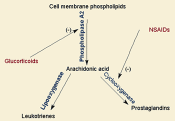
Posted in General.

Tagged with , , , , , , , , , , , , , , , , , , , , , , , , , , , , .