Categories:

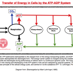
Transfer of Energy in Cells by the ATP-ADP System

Also see:
Energy Flow: Plant World and Animal World
The Sun: Source of All Biological Energy
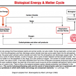
Biological Energy & Matter Cycle
Collection of FPS Charts
Cellular Energy Production – Aerobic Respiration – The Krebs Cycle
Promoters of Efficient v. Inefficient Metabolism
Comparison: Oxidative Metabolism v. Glycolytic Metabolic
Comparison: Carbon Dioxide v. Lactic Acid
Carbon Dioxide Basics
Protect the Mitochondria

Posted in General.


Biological Energy & Matter Cycle

Also see:
Energy Flow: Plant World and Animal World
The Sun: Source of All Biological Energy
Transfer of Energy in Cells by the ATP-ADP System
Collection of FPS Charts
Cellular Energy Production – Aerobic Respiration – The Krebs Cycle
Promoters of Efficient v. Inefficient Metabolism
Comparison: Oxidative Metabolism v. Glycolytic Metabolic
Comparison: Carbon Dioxide v. Lactic Acid
Carbon Dioxide Basics
Protect the Mitochondria

Posted in General.


The Sun: Source of All Biological Energy

Also see:
Biological Energy & Matter Cycle
Energy Flow: Plant World and Animal World
Transfer of Energy in Cells by the ATP-ADP System
Collection of FPS Charts
Promoters of Efficient v. Inefficient Metabolism
Comparison: Oxidative Metabolism v. Glycolytic Metabolic
Comparison: Carbon Dioxide v. Lactic Acid
Carbon Dioxide Basics
Protect the Mitochondria
Light is Right
Using Sunlight to Sustain Life

The sun is the source of all biological energy, providing plants the means to produce glucose which in turn provides the animals that eat the plants the energy needed to perform biological work.

Posted in General.


Energy Flow: Plant World and Animal World

Also see:
The Sun: Source of All Biological Energy
Biological Energy & Matter Cycle
Transfer of Energy in Cells by the ATP-ADP System
Collection of FPS Charts
Cellular Energy Production – Aerobic Respiration – The Krebs Cycle
Promoters of Efficient v. Inefficient Metabolism
Comparison: Oxidative Metabolism v. Glycolytic Metabolic
Comparison: Carbon Dioxide v. Lactic Acid
Carbon Dioxide Basics
Protect the Mitochondria

A look at the energy “balance sheets” of the animal and plant world reveals that they are nearly the inverse of each other. Photosynthesis is the reversal of cellular respiration and vice versa.

Posted in General.


PUFA, Ketones, and Sugar Restriction Promote Tumor Growth

Also see:
How cancer cells rewire their metabolism to survive
Tumor Bearing Organisms – Lipolysis and Ketogenesis as Signs of Chronic Stress
Fatty Acid Synthase (FAS), Vitamin D, and Cancer
Free Fatty Acids Suppress Cellular Respiration
PUFA Promote Cancer
PUFA Decrease Cellular Energy Production
Israeli Paradox: High Omega -6 Diet Promotes Disease
Low Blood Sugar Basics
Dietary PUFA Reflected in Human Subcutaneous Fat Tissue
Towards a morphogenetic perspective on cancer
Israeli Paradox: High Omega -6 Diet Promotes Disease
Toxicity of Stored PUFA
PUFA Accumulation & Aging
The Randle Cycle
Maternal PUFA Intake Increases Breast Cancer Risk in Female Offspring
Maternal Ingestion of Tryptophan and Cancer Risk in Female Offspring
Polyamines and Cancer
Carrageenan, Inflammation, Cancer, Immunity
AIDS patients – High PUFA and High Cortisol in Blood
Cancer Cells Are What They Eat, and It’s Not Glucose
Sugar Feeds Thyroid

https://www.youtube.com/watch?v=S8o8GvyGRYc

Quotes by Ray Peat, PhD:
“Starving for sugar causes the body to break down proteins to make glucose, weakening the organism’s resistance, providing amino acids for tumor growth, and probably providing ammonia and other things that stimulate growth and interfere with differentiation. The liberated free fatty acids in either sugar deprivation or diabetes (in which cells are starved for glucose) stimulate tumor growth.”

“There are many people currently recommending fish oil (or other highly unsaturated oils) for preventing or treating cancer, and it has become almost as common to recommend a sugar free diet, “because sugar feeds cancer.” This is often, incorrectly, said to be the meaning of Warburg’s demonstration that cancer cells have a respiratory defect that causes them to produce lactic acid from glucose even in the presence of oxygen. Cancer cells use glucose and the amino acid glutamine primarily for synthetic purposes, and use fats as their energy source;the growth stimulating effect of the “essential fatty acids” (Sueyoshi and Nagao, 1962a; Holley, et al., 1974) shows that depriving a tumor of those fats retards its growth. The great energetic inefficiency of the cancer metabolism, which causes it to produce a large amount of heat and to cause systemic stress, failure of immunity, and weight loss, is because it synthesizes fat from glucose and amino acids, and then oxidizes the fat as if it were diabetic.”

“”Cancer metabolism” or stress metabolism typically involves an excess of the adaptive hormones, resulting from an imbalance of the demands made on the organism and the resources available to the organism. Excessive stimulation depletes glucose and produces lactic acid, and causes cortisol to increase, causing a shift to the consumption of fat and protein rather than glucose. Increased cortisol activates the Randle effect (the inhibition of glucose oxidation by free fatty acids), accelerates the breakdown of protein into amino acids, and activates the enzyme fatty acid synthase, which produces fatty acids from amino acids and pyruvate, to be oxidized in a “futile cycle,” producing heat, and increasing the liberation of ammonia from the amino acids. Ammonia suppresses respiratory, and stimulates glycolytic, activity.”

“In Warburg’s view, cancer has a unique requirement for glucose, because of its “respiratory defect.” The ability of cancer to consume large amounts of glucose means that the body interprets cancer as stress, since the waste of glucose is the key feature of stress. The body’s response to decreasing glucose is to produce larger amounts of adrenal hormones, especially cortisol. Cortisol raises the blood glucose supply by converting tissue proteins into sugar. This hormonal response to the sugar appetite of cancer is the main cause of the wasting syndrome which makes cancer so serious, and which destroys the thymus gland, causing the immune system to lose its guidance. V. S. Shapot’s work, showing the involvement of glucose and cortisol in cancer, provides part of the context for understanding the implications of Warburg’s view of cancer as a “respiratory defect.””

“Ketones are very protective as a fuel, but the problem is that they are produced as a result of metabolic stress. If the liver is extremely good, it can store enough glycogen for a day, but chronic, frequent, stress usually damages the liver’s ability to store glycogen.”

Cancer Res. 1987 Feb 15;47(4):1065-8.
Blood nutrient concentrations and tumor growth in vivo in rats: relationships during the onset of an acute fast.
Sauer LA, Dauchy RT.
The rate of tumor growth in vivo in adult rats (250- to 350-g total body weight) is stimulated during an acute fast. No tumor growth stimulation is observed in fasted immature rats (less than about 200-g total body weight). The different tumor growth responses in rats of these two age groups appear to depend on the increased availability to the tumor of nutrients from host fat stores in adult rats. Immature rats, which lack significant fat stores, show neither hyperlipemia nor ketosis during fasting. These experiments were performed to determine the relationship between blood fat store-derived nutrient concentrations and the onset of stimulated tumor growth in fasted adult rats. Animals were matched for tumor size and growth during a period of ad libitum feeding preceding the fast. Tumor growth was documented by increased size and incorporation of [methyl-3H]thymidine into tumor DNA. Mobilization of host fat stores leading to increased blood concentrations of free fatty acids, glycerol, ketone bodies, and triglycerides started about 7 h after food was removed and reached its maximum after about 15 h. Increased rates of tumor growth and incorporation of thymidine into tumor DNA correlated closely with the higher circulating nutrient concentrations. Both the nutrient concentrations and tumor growth were decreased by refeeding. These findings suggest that the availability of nutrients derived from host fat stores may be rate limiting for tumor growth in vivo.

Cancer Res. 1986 Jul;46(7):3469-75.
Stimulation of tumor growth in adult rats in vivo during an acute fast.
Sauer LA, Nagel WO, Dauchy RT, Miceli LA, Austin JE.
These experiments investigate an increase in tumor growth that occurs in adult rats in vivo during an acute fast. The effects of feeding, fasting, and underfeeding on the growth of Morris hepatomas 5123C and 7288CTC in Buffalo rats and of Walker carcinoma 256 and Jensen sarcoma in Sprague-Dawley rats were studied. Animals were matched for tumor size and growth during a period of ad libitum feeding preceding the fasting or underfeeding. Tumor growth was documented by increased size and incorporation of [methyl-3H]thymidine into tumor DNA. Fasting increased the rate of growth of the tumors 3 to 4 times over that measured in fed rats. This effect began during the first day of fasting and ended abruptly on refeeding. After refeeding tumor growth slowed to the rate in fed rats. Tumors from fed or fasted rats were not different in cellularity or dry weight/g wet weight. A positive growth response in the tumor required lipolysis and ketosis in the host. No stimulation was observed during an acute fast in either immature rats or in mature rats whose weights had been reduced by underfeeding. These animals have small fat stores and show no increase in arterial blood free fatty acid or ketone body concentrations during an acute fast. Finally, underfeeding of adult rats raised the blood concentrations of these nutrients to values that were intermediate between those in fasted and fed rats. Tumor growth rates in these rats were intermediate between those in fasted and fed rats. The results support the proposal that an increase in availability of free fatty acids and/or ketone bodies is the stimulus that increases the rate of tumor growth during an acute fast.

Cancer Res. 1987 Apr 1;47(7):1756-61.
Stimulation of tumor growth in adult rats in vivo during acute streptozotocin-induced diabetes.
Sauer LA, Dauchy RT
The effects of acute diabetes mellitus on the growth of Morris hepatoma 7288CTC and Jensen sarcoma were studied in fed, young (less than 200 g), and adult (greater than 250 g) rats. Animals were matched for tumor size and growth; the rates of tumor growth were the same in fed, young and adult nondiabetic rats. Diabetes was induced by the i.v. injection of streptozotocin (65 mg/kg total body weight) into tumor-bearing rats and changes in arterial blood nutrient concentrations were compared to changes in the rates of tumor growth and DNA synthesis. In young rats acute diabetes did not increase the blood concentrations of the fat store-derived nutrients and did not increase the rate of tumor growth. In adult rats, however, acute diabetes raised the arterial blood free fatty acid, glycerol, triglyceride, and ketone body concentrations to high levels and increased the rate of tumor growth about three times over that observed in untreated rats. Progress curves for the mobilization of host fat stores and for incorporation of [methyl-3H]thymidine into tumor DNA during the onset of diabetes showed that these activities were closely correlated in adult rats. Both processes began to increase 2 to 4 h after streptozotocin treatment, reached an initial peak at 12 to 16 h, decreased to a low point at 18 to 20 h, and then increased again to the new steady state after 23 to 24 h. The results indicate that the rate of tumor growth in rats in vivo is limited by the availability of a substance(s) present in the hyperlipemic blood of adult diabetic rats. The tight relationship between host lipolysis and tumor growth suggests that the substance(s) is derived from host fat stores.

Cancer Res. 1988 Jun 1;48(11):3106-11.
Identification of linoleic and arachidonic acids as the factors in hyperlipemic blood that increase [3H]thymidine incorporation in hepatoma 7288CTC perfused in situ.
Sauer LA, Dauchy RT.
Tumor growth and the incorporation of [3H]thymidine into tumor DNA in vivo are increased about 3 times in adult rats (greater than 250 g) after 1 to 2 days of starvation or the induction of diabetes with streptozotocin. These tumor growth responses require hyperlipemia and are reversed by refeeding or insulin treatment, respectively. They do not occur in young tumor-bearing rats (less than about 150 g) that lack appreciable fat stores. A direct relationship between the increased rates of both [3H]thymidine incorporation and tumor growth and host hyperlipemia suggests that tumor cell renewal in vivo in fed rats is limited by substances that are present in hyperlipemic blood. In this study we used a procedure for perfusion of solid tumors in situ to measure the sensitivity of tumor [3H]thymidine incorporation to hyperlipemic blood and to identify the rate-limiting substances. Tissue-isolated Morris hepatomas (7288CTC) growing in young or adult Buffalo rats were perfused with blood from donor rats. Hyperlipemic blood for perfusion was obtained from 2-day starved tumor-bearing (Buffalo) or non-tumor-bearing (Buffalo or Lewis) rats. At the end of the perfusions the tumors were labeled with a pulse of [3H]thymidine (2 microCi/g estimated tumor wet weight). [3H]Thymidine incorporation in tumors growing in fed adult rats was increased from 80 +/- 5 (SD) dpm/micrograms DNA at zero time (before perfusion) to 209 +/- 9 dpm/micrograms DNA (n = 3) after perfusion for 3 h. Tumors growing in fed or starved young rats showed similar responses, and hyperlipemic blood from non-tumor-bearing rats was as effective as hyperlipemic blood from tumor-bearing rats. Perfusion of tumors growing in starved rats with normolipemic blood from fed adult rats decreased [3H]thymidine incorporation from 211 +/- 13 dpm/micrograms DNA before perfusion to 68 +/- 9 dpm/micrograms DNA (n = 3) after perfusion for 3 h. Cells, plasma, and plasma subfractions from hyperlipemic blood were reconstituted to whole blood using plasma, cells, and whole blood, respectively, from fed rats and the mixtures were perfused into tumors growing in fed adult rats. Mixtures containing hyperlipemic plasma, lipid extracts (ethanol:acetone, 1:1) of hyperlipemic plasma, or albumin from hyperlipemic plasma increased tumor [3H]thymidine incorporation. Free fatty acid concentrations were increased about five times in hyperlipemic plasma and perfusion of tumors with normolipemic blood containing added linoleic and arachidonic acids increased [3H]thymidine incorporation. Blood mixtures containing palmitic, stearic, and oleic acids were inactive.(ABSTRACT TRUNCATED AT 400 WORDS)

J Nutr. 1997 Jul;127(7):1412-21.
Dietary linoleic acid intake controls the arterial blood plasma concentration and the rates of growth and linoleic acid uptake and metabolism in hepatoma 7288CTC in Buffalo rats.
Sauer LA, Dauchy RT, Blask DE.
In this study, we tested the hypothesis that dietary linoleic acid intake controls the arterial blood plasma linoleic acid concentration and the rates of tumor growth and linoleic acid metabolism in vivo. Seven groups of young male Buffalo rats (11-21 rats/group) were given free access to semipurified diets containing different amounts of corn and/or olive oils. Four other groups (7-11 rats/group) were 30% energy-restricted. Each experiment included periods for rat growth and plasma lipid stabilization (6 wk), measurement of mean daily arterial blood plasma fatty acid concentrations (3 wk), surgical implantation of a subcutaneous tissue-isolated hepatoma 7288CTC, tumor growth and harvest (2-4 wk). Linoleic + arachidonic acid (P = 0.007) and oleic acid (P = 0.002) concentrations in arterial blood plasma were increased as dietary intake of linoleic and oleic acids was increased, respectively. In rats given free access to food, tumor growth was directly dependent on the plasma concentrations of linoleic (P < 0.001) and arachidonic acids (P = 0.04). Tumor growth in energy-restricted rats was dependent only on the linoleic acid concentration (P = 0.008). Energy restriction itself caused a growth inhibition independent of plasma linoleic acid. The linoleic acid and total fatty acid concentrations of tumor triacylglycerols were directly dependent on the plasma linoleic acid concentration in rats given free access to food (P = 0.009). Hepatoma 7288CTC (both in vivo and during perfusion in situ) supported a dose-dependent conversion (P < 0.001) of plasma linoleic acid to the mitogen, 13-hydroxy-9, 11-octadecadienoic acid. We conclude that increased arterial blood plasma linoleic acid concentrations, caused by increased dietary intakes, specifically stimulate growth, lipid storage and linoleic acid metabolism in hepatoma 7288CTC in vivo.

Cancer Res. 1983 Aug;43(8):3497-503.
Ketone body, glucose, lactic acid, and amino acid utilization by tumors in vivo in fasted rats.
Sauer LA, Dauchy RT.
Arteriovenous differences for acetoacetate, beta-hydroxybutyrate, glucose, lactic acid, and glutamine and other amino acids were measured across Morris hepatomas 5123C, 7777, and 7288CTCF and Walker sarcocarcinoma 256 in vivo in rats fasted for 2 days. The acetoacetate and beta-hydroxybutyrate concentrations in arterial whole blood of fasted tumor-bearing rats were 0.52 +/- 0.06 and 1.82 +/- 0.19 mM (S.E., n = 38), respectively. Both ketone bodies were utilized by the tumors, and the rates of utilization were directly related to the rates of supply. The mean utilization rates for acetoacetate and beta-hydroxybutyrate were 13.9 +/- 2.9 (range, 0 to 64; n = 30) and 24.7 +/- 4.4 (range, 0 to 145; n = 38) nmol/min/g tumor wet weight, respectively. Eight of the tumors produced acetoacetate, presumably from utilized beta-hydroxybutyrate. An average of 52% of the acetoacetate and 30% of the beta-hydroxybutyrate carried in the arterial blood was removed during one pass through the tumors. The concentrations of glucose and glutamine in the arterial whole blood of fasted tumor-bearing rats (n = 38) were 6.55 +/- 0.3 and 0.76 +/- 0.02 mM, respectively; both of these substrates were utilized at rates that were directly proportional to the rates of supply. The mean rates of glucose and glutamine utilization for all tumors in fasted rats were 101 +/- 11 (range, 3 to 313) and 8.2 +/- 1.1 (range, 0 to 25.1) nmol/min/g tumor wet weight, respectively. Thirty-six % of the glucose and 25% of the glutamine supplied to the tumors was utilized. Comparison (by linear regression and analysis of covariance) of the rates of supply and utilization of glucose and glutamine in tumors growing in fasted versus fed rats indicated that these substrates are utilized more efficiently by tumors growing in fasted animals. Lactic acid was either produced or utilized, depending on the arterial whole-blood concentration. Production or utilization occurred, respectively, when the arterial lactate concentration was less or greater than 1 to 3 mM. The arterial whole-blood amino acids (except glutamine) were utilized at rates that ranged from 1 to 4 nmol/min/g tumor wet weight. The results indicate that energy production for tumor growth in fasted rats is supported, in part, by an increased availability of ketone bodies, by an increased efficiency of utilization of glucose and glutamine, and, under certain circumstances, by utilization of lactic acid.

Cell Cycle. 2011 Apr 15;10(8):1271-86. doi: 10.4161/cc.10.8.15330.
Ketones and lactate increase cancer cell “stemness,” driving recurrence, metastasis and poor clinical outcome in breast cancer: achieving personalized medicine via Metabolo-Genomics.
Martinez-Outschoorn UE, Prisco M, Ertel A, Tsirigos A, Lin Z, Pavlides S, Wang C, Flomenberg N, Knudsen ES, Howell A, Pestell RG, Sotgia F, Lisanti MP.
Previously, we showed that high-energy metabolites (lactate and ketones) “fuel” tumor growth and experimental metastasis in an in vivo xenograft model, most likely by driving oxidative mitochondrial metabolism in breast cancer cells. To mechanistically understand how these metabolites affect tumor cell behavior, here we used genome-wide transcriptional profiling. Briefly, human breast cancer cells (MCF7) were cultured with lactate or ketones, and then subjected to transcriptional analysis (exon-array). Interestingly, our results show that treatment with these high-energy metabolites increases the transcriptional expression of gene profiles normally associated with “stemness,” including genes upregulated in embryonic stem (ES) cells. Similarly, we observe that lactate and ketones promote the growth of bonafide ES cells, providing functional validation. The lactate- and ketone-induced “gene signatures” were able to predict poor clinical outcome (including recurrence and metastasis) in a cohort of human breast cancer patients. Taken together, our results are consistent with the idea that lactate and ketone utilization in cancer cells promotes the “cancer stem cell” phenotype, resulting in significant decreases in patient survival. One possible mechanism by which these high-energy metabolites might induce stemness is by increasing the pool of Acetyl-CoA, leading to increased histone acetylation, and elevated gene expression. Thus, our results mechanistically imply that clinical outcome in breast cancer could simply be determined by epigenetics and energy metabolism, rather than by the accumulation of specific “classical” gene mutations. We also suggest that high-risk cancer patients (identified by the lactate/ketone gene signatures) could be treated with new therapeutics that target oxidative mitochondrial metabolism, such as the anti-oxidant and “mitochondrial poison” metformin. Finally, we propose that this new approach to personalized cancer medicine be termed “Metabolo-Genomics,” which incorporates features of both 1) cell metabolism and 2) gene transcriptional profiling. Importantly, this powerful new approach directly links cancer cell metabolism with clinical outcome, and new therapeutic strategies for inhibiting the TCA cycle and mitochondrial oxidative phosphorylation in cancer cells.

Cancer. 1988 Feb 15;61(4):689-701.
Peripheral blood lymphocytes from patients with cancer lack interleukin-2 receptors.
Hakim AA.
When tumor cells develop in healthy adults, they activate the cellular immune system–natural killer (NK) cells, antigen-specific cytotoxic lymphocytes (CTL), and the synthesis of antigen specific cytotoxic antibodies. These are aimed at killing the intruding cells. However, in cancer patients the tumor continues to grow. As tumor cells proliferate, they were shown to release factors that mediate the inactivation of the host immune defense systems. The study documented in this article examined peripheral blood lymphocytes, mononuclear cells (MNC), NK cells, T-helper cells (THC). This study confirmed the interaction of the released inhibitor factors with these mononuclear cells. NULL cells from healthy adults responding to interleukin-2 (IL-2) and NILL cells from patients with metastatic breast carcinoma nonresponsive to IL-2 were also isolated by the standard antibodies-pinning technique. The cells were obtained from age-matched subjects: ten healthy adults; ten patients each from Stage I, II, III, and IV metastatic breast carcinoma (BCa-I, BCa-II, BCa-III, and BCa-IV or MBCa); and ten patients with benign breast disease (BBD). The responsiveness of these THC, PBMNC, NK, NULL, and NILL cells in vitro to graded levels of phytohemagglutinin (PHA), Concanavalin A (Con A), and recombinant interleukin-2 (rIL-2) was examined. Responsiveness was monitored by 3H-thymidine (3H-TdR) uptake, production and release of IL-2, interleukin-2 receptor (IL-2R), and cytotoxic activities against K-562 cells and breast carcinoma short-term cell lines. A lack of functional IL-2R in peripheral blood lymphocytes from patients with metastatic breast carcinoma was confirmed by nonsignificant anti-Tac antibody binding. An elevation in the expression of cell surface antigen GP-120 has been observed to be associated with the activation in vitro of T-cells from healthy adults and from patients with benign breast disease, but not of T-cells from patients with breast carcinoma. Biochemical studies of the GP-120 using high performance liquid chromatography combined with nitrocellulose blotting confirmed that the glycoprotein was resistant to trypsin and chymotrypsin, but susceptible to pronase. It contained sialic acid and lactosaminoglycan as O-linked sugars. It could be labeled with pariodate/NaB(3H4) and is recognized by MAbT-305 monoclonal antibodies. It contained sialic acid linked (2—3) to galactose.(ABSTRACT TRUNCATED AT 400 WORDS)

When cancer cells are implanted into a healthy host, they seldom grow, but when implanted into a host who already has cancer, they grow. (Hakim, 1988.) The healthy host provides many restorative factors, the sick host provides additional harmful factors, but few restorative factors (Chekulaev, et aI., 1987.) -Ray Peat, PhD

Biokhimiia. 1987 Sep;52(9):1501-11.
[Activation of lipolysis and ketogenesis in tumor-bearing animals as a reflection of chronic stress states].
[Article in Russian]
Chekulaev VA, Shelepov VP, Pasha-zade GR, Shapot VS.
In order to elucidate the peculiarities of brain metabolism in tumour-bearing organisms, the arterio-venous (A-V) content of glucose, acetoacetate (Ac-Ac), beta-hydroxybutyrate (beta-HB) and non-esterified fatty acids (NEFA) in growing Zajdela ascite hepatoma (ZAH) and solid hepatoma 27 (H-27) was compared. Analysis of metabolic patterns of healthy, starving and fed recipients (ZAH and H-27) revealed the inadequacy of the concepts on anorexia as being the cause of carbohydrate-lipid metabolic disturbances. In tumour-bearing organisms lipolysis and ketogenesis reflect the tumour-induced chronic stress. Absorption of beta-HB and release of Ac-Ac by brain were observed at all stages of malignant growth. This is probably due to a partial switch-over of brain metabolism to non-carbohydrate energy sources. Besides, certain stages of tumour growth are associated with active assimilation of NEFA by brain. A correlation between the A-V difference with respect to glucose and Ac-Ac as well as between the glucose and NEFA contents was established. It was assumed that the A-V difference in glucose is the main regulator of ketone body metabolism.

Prog Clin Biol Res. 1983;111:89-109.
Energy metabolism in trauma and sepsis: the role of fat.
Wolfe RR, Shaw JH, Durkot MJ.
There seems little doubt that there are signals for the increased mobilization of fat in shock, trauma, and sepsis. Whether those signals are reflected by an actual increase in mobilization is dependent on many variables including cardiovascular status. A hypothetical scheme based on our own experiments in the hyperdynamics phases of response to burn injury and to sepsis is presented in Figure 8. According to this scheme, catecholamines stimulate lipolysis in the adipose tissue, resulting in the release of glycerol and FFA into the plasma at increased rates. The glycerol is cleared by the liver and converted into glucose–a process stimulated by, among other things, glucagon. Some of the increased flux of FFA is also cleared by the liver, whereupon the fatty acids are incorporated into VLDL and released again into the plasma. The increased FFA levels also exert a dampening effect on the factors stimulating hepatic glucose production. At the periphery, plasma FFA as well as VLDL fatty acids are taken up at an increased rate. The tissues are attuned to the oxidation of fat, and as a consequence most of the energy production is derived from fat oxidation. The increased fatty acids exert an inhibitory effect on the complete oxidation of glucose, so although glucose may be taken up at an accelerated rate, the relative contribution of glucose oxidation to total energy production may fall. Rather than being completely oxidized, pyruvate is reduced to lactate and released into the plasma at an accelerated rate. The lactate then contributes to the production of glucose in the liver, completing a cyclical process called the Cori Cycle. Although all aspects of this scheme are supported by data highlighted in this paper, it certainly must be an oversimplification of the overall response of substrate metabolism to trauma and sepsis. It is presented for the purpose of highlighting the potential role of fat as a controller of the metabolic response, and to suggest that the enhanced mobilization and oxidation of fat is one of the fundamental responses to stress.

Cell Cycle. 2010 Sep 1;9(17):3506-14. Epub 2010 Sep 21.
Ketones and lactate “fuel” tumor growth and metastasis: Evidence that epithelial cancer cells use oxidative mitochondrial metabolism.
Bonuccelli G1, Tsirigos A, Whitaker-Menezes D, Pavlides S, Pestell RG, Chiavarina B, Frank PG, Flomenberg N, Howell A, Martinez-Outschoorn UE, Sotgia F, Lisanti MP.
Previously, we proposed a new model for understanding the “Warburg effect” in tumor metabolism. In this scheme, cancer-associated fibroblasts undergo aerobic glycolysis and the resulting energy-rich metabolites are then transferred to epithelial cancer cells, where they enter the TCA cycle, resulting in high ATP production via oxidative phosphorylation. We have termed this new paradigm “The Reverse Warburg Effect.” Here, we directly evaluate whether the end-products of aerobic glycolysis (3-hydroxy-butyrate and L-lactate) can stimulate tumor growth and metastasis, using MDA-MB-231 breast cancer xenografts as a model system. More specifically, we show that administration of 3-hydroxy-butyrate (a ketone body) increases tumor growth by ∼2.5-fold, without any measurable increases in tumor vascularization/angiogenesis. Both 3-hydroxy-butyrate and L-lactate functioned as chemo-attractants, stimulating the migration of epithelial cancer cells. Although L-lactate did not increase primary tumor growth, it stimulated the formation of lung metastases by ∼10-fold. Thus, we conclude that ketones and lactate fuel tumor growth and metastasis, providing functional evidence to support the “Reverse Warburg Effect”. Moreover, we discuss the possibility that it may be unwise to use lactate-containing i.v. solutions (such as Lactated Ringer’s or Hartmann’s solution) in cancer patients, given the dramatic metastasis-promoting properties of L-lactate. Also, we provide evidence for the up-regulation of oxidative mitochondrial metabolism and the TCA cycle in human breast cancer cells in vivo, via an informatics analysis of the existing raw transcriptional profiles of epithelial breast cancer cells and adjacent stromal cells. Lastly, our findings may explain why diabetic patients have an increased incidence of cancer, due to increased ketone production, and a tendency towards autophagy/mitophagy in their adipose tissue.

Brain Dev. 2010 Apr;32(4):318-22. doi: 10.1016/j.braindev.2009.04.009. Epub 2009 May 13.
Tumor growth in patients with tuberous sclerosis complex on the ketogenic diet.
Chu-Shore CJ1, Thiele EA.
PURPOSE:
New evidence is emerging that the availability of nutrients plays a key role in regulating the mammalian target of rapamycin complex-1 (mTORC1) signaling pathway in human cancers. Tuberous sclerosis complex (TSC) is a genetic disorder which results in the growth of hamartomatous lesions in multiple organs due to insufficient suppression of the mTORC1 pathway. A minority of patients with TSC who develop epilepsy which is intractable to standard anticonvulsant medical and/or surgical treatments are treated with the ketogenic diet. To provide insight into the effects of nutrient manipulation on tumor growth in this condition, we describe our experience in a unique group of patients with known tuberous sclerosis complex who are on the ketogenic diet for seizure control.
METHODS:
A retrospective chart review was performed of patients with TSC treated with the ketogenic diet between January 2002 and May 2007 at Massachusetts General Hospital.
RESULTS:
Five patients with definite TSC underwent serial imaging for tumor growth while on the ketogenic diet or had unchanged imaging prior to the onset of the diet and after termination. Three out of five patients, all children, had progression of a known tumor or tumors or the development of a new tumor while on the ketogenic diet.
CONCLUSION:
In this limited case series of five TSC patients, the ketogenic diet did not induce tumor regression or suppress the growth of TSC-related tumors.

Cell Cycle. 2012 Nov 1;11(21):3964-71. doi: 10.4161/cc.22137. Epub 2012 Sep 19.
Ketone body utilization drives tumor growth and metastasis.
Martinez-Outschoorn UE, Lin Z, Whitaker-Menezes D, Howell A, Sotgia F, Lisanti MP.
We have previously proposed that catabolic fibroblasts generate mitochondrial fuels (such as ketone bodies) to promote the anabolic growth of human cancer cells and their metastasic dissemination. We have termed this new paradigm “two-compartment tumor metabolism.” Here, we further tested this hypothesis by using a genetic approach. For this purpose, we generated hTERT-immortalized fibroblasts overexpressing the rate-limiting enzymes that promote ketone body production, namely BDH1 and HMGCS2. Similarly, we generated MDA-MB-231 human breast cancer cells overexpressing the key enzyme(s) that allow ketone body re-utilization, OXCT1/2 and ACAT1/2. Interestingly, our results directly show that ketogenic fibroblasts are catabolic and undergo autophagy, with a loss of caveolin-1 (Cav-1) protein expression. Moreover, ketogenic fibroblasts increase the mitochondrial mass and growth of adjacent breast cancer cells. However, most importantly, ketogenic fibroblasts also effectively promote tumor growth, without a significant increase in tumor angiogenesis. Finally, MDA-MB-231 cells overexpressing the enzyme(s) required for ketone re-utilization show dramatic increases in tumor growth and metastatic capacity. Our data provide the necessary genetic evidence that ketone body production and re-utilization drive tumor progression and metastasis. As such, ketone inhibitors should be designed as novel therapeutics to effectively treat advanced cancer patients, with tumor recurrence and metastatic disease. In summary, ketone bodies behave as onco-metabolites, and we directly show that the enzymes HMGCS2, ACAT1/2 and OXCT1/2 are bona fide metabolic oncogenes.

Cell Cycle. 2012 Nov 1; 11(21): 3956–3963.
Ketone bodies and two-compartment tumor metabolism: Stromal ketone production fuels mitochondrial biogenesis in epithelial cancer cells
Ubaldo E. Martinez-Outschoorn, Zhao Lin, Diana Whitaker-Menezes, Anthony Howell, Michael P. Lisanti, and Federica Sotgia
We have previously suggested that ketone body metabolism is critical for tumor progression and metastasis. Here, using a co-culture system employing human breast cancer cells (MCF7) and hTERT-immortalized fibroblasts, we provide new evidence to directly support this hypothesis. More specifically, we show that the enzymes required for ketone body production are highly upregulated within cancer-associated fibroblasts. This appears to be mechanistically controlled by the stromal expression of caveolin-1 (Cav-1) and/or serum starvation. In addition, treatment with ketone bodies (such as 3-hydroxy-butyrate, and/or butanediol) is sufficient to drive mitochondrial biogenesis in human breast cancer cells. This observation was also validated by unbiased proteomic analysis. Interestingly, an MCT1 inhibitor was sufficient to block the onset of mitochondrial biogenesis in human breast cancer cells, suggesting a possible avenue for anticancer therapy. Finally, using human breast cancer tumor samples, we directly confirmed that the enzymes associated with ketone body production (HMGCS2, HMGCL and BDH1) were preferentially expressed in the tumor stroma. Conversely, enzymes associated with ketone re-utilization (ACAT1) and mitochondrial biogenesis (HSP60) were selectively associated with the epithelial tumor cell compartment. Our current findings are consistent with the “two-compartment tumor metabolism” model. Furthermore, they suggest that we should target ketone body metabolism as a new area for drug discovery, for the prevention and treatment of human cancers.

Front. Endocrinol., 02 February 2016
Fenofibrate Induces Ketone Body Production in Melanoma and Glioblastoma Cells
Maja M. Grabacka, Anna Wilk, Anna Antonczyk, Paula Banks, Emilia Walczyk-Tytko, Matthew Dean, Malgorzata Pierzchalska, and Krzysztof Reiss
“The capacity of cancer cells to produce ketone bodies described here is quite different from previous reports, including series of papers (58–60) by the Lisanti group and coworkers regarding the possibility of ketogenesis in stromal fibroblasts surrounding breast cancer lesions. In those papers, the authors showed that cancer cells actively contributed to the stromal fibroblasts’ metabolic reprograming and took advantage of the subsequent ketone body consumption for energy generation. This is a special property of epithelia-derived tumors, such as breast carcinomas, since other cancer types do not express the proper enzymatic machinery for ketone body utilization as a source of energy (25, 28, 34). More often, cancers such as neuroblastomas or astrocytomas do absorb ketone bodies, but they use them for lipid synthesis rather than energy production (61–63).”
“In conclusion, it is quite surprising that fenofibrate is able to reprogram melanoma and glioblastoma metabolic pathways in such a way that they suffer from an energy deficit but are still forced to produce ketone bodies. This finding is important because, as far as we are aware, ketogenesis in cancer cells has not been described before and the underlying mechanism still waits to be revealed. The ketone bodies released by glioblastoma cells could serve not only as a fuel but also as a cytoprotective signaling molecule for neurons in the microenvironment surrounding the tumor tissue.”

Posted in General.

Tagged with , , , , , , , , , , , , , , , , , .


Body Temperature, Metabolism, and Obesity

Also see:
Ray Peat, PhD on Thyroid, Temperature, Pulse, and TSH
Temperature and Pulse Basics & Monthly Log
Thyroid, Temperature, Pulse
Is 98.6 Really Normal?
Metabolism, Brain Size, and Lifespan in Mammals
Promoters of Efficient v. Inefficient Metabolism
Inflammation from Decrease in Body Temperature
Melatonin Lowers Body Temperature
Menopausal Estrogen Therapy Lowers Body Temperature
Thyroid Function, Pulse Rate, & Temperature
“Curing” a High Metabolic Rate with Unsaturated Fats
Fat Deficient Animals – Activity of Cytochrome Oxidase

Metabolism. 2009 Jun;58(6):871-6. doi: 10.1016/j.metabol.2009.02.017.
Is obesity associated with lower body temperatures? Core temperature: a forgotten variable in energy balance. (full paper)
Landsberg L, Young JB, Leonard WR, Linsenmeier RA, Turek FW.
The global increase in obesity, along with the associated adverse health consequences, has heightened interest in the fundamental causes of excessive weight gain. Attributing obesity to “gluttony and sloth”, blaming the obese for overeating and limiting physical activity, oversimplifies a complex problem, since substantial differences in metabolic efficiency between lean and obese have been decisively demonstrated. The underlying physiological basis for these differences have remained poorly understood. The energetic requirements of homeothermy, the maintenance of a constant core temperature in the face of widely divergent external temperatures, accounts for a major portion of daily energy expenditure. Changes in body temperature are associated with significant changes in metabolic rate. These facts raise the interesting possibility that differences in core temperature may play a role in the pathophysiology of obesity. This review explores the hypothesis that lower body temperatures contribute to the enhanced metabolic efficiency of the obese state.

Some quotables:

Resting (or “basal”) metabolic rate (RMR) accounts for
approximately 80%of energy output. About two thirds of RMR
is for maintenance of homeothermy (warm-bloodedness); about
one third is to maintain cellular integrity, ionic gradients, protein
turnover, and the like [6-8] Resting metabolic rate is largely
regulated by thyroid hormones, with a minor contribution from
the sympathetic nervous system.
Resting metabolic rate differs
by as much as 600 kcal/d for a 70-kg man [8].
Physical activity (exercise) accounts for about 10% in
truly sedentary humans; in addition to intentional activity,
this category includes nonpurposeful motion such as
fidgeting, which may differ among lean and obese
individuals [9], as well as upright posture [10].
The remaining 10% is frequently referred to as thermogenesis,
which means heat production unrelated to physical
activity.

It should be emphasized that, for nonsedentary individuals,
the activity component may be much greater than 10%
of total energy expenditure. Evidence has been developed
indicating that the combination of activity plus adaptive
thermogenesis accounts for about 44% of total energy
expenditure on average, meaning that RMR would constitute
about 56% of total energy expenditure in normally active
humans [15], as compared with 80% in the truly sedentary.

A lesser ability to dissipate ingested calories is one
example of a thrifty metabolic trait that has evolved to
promote survival in the face of fluctuations in food
availability
. Since the initial formulation of the “thrifty
gene” hypothesis by James Neel in 1962 [16], the nature of
thrifty traits has been the subject of considerable research
and speculation. A recent formulation [17] highlights 2
distinct components: (1) decreased metabolic rate and/or a
diminished capacity for “thermogenesis” and (2) decreased
insulin sensitivity. These 2 components address the 2 main
physiologic imperatives of starvation: energy conservation
and protein preservation. A decrease in metabolic rate would
lead to more efficient storage of calories as fat, thereby
prolonging survival during famine; during periods of
abundance and in the face of dietary excess, this trait
would predispose to obesity. Resistance to the action of
insulin would divert glucose from skeletal muscle, which can
use fat-derived substrates, to the brain, an organ almost
entirely obligated to the use of glucose. In the presence of
famine, insulin resistance would spare muscle breakdown by
lessening the need for gluconeogenesis from protein; in the
face of an abundant food supply, however, and in association
with dietary excess, insulin resistance would predispose to
type 2 diabetes mellitus.

“Both metabolic efficiency and insulin resistance, moreover,
are known to vary among different individuals in the
same population. The survival value of these thrifty traits,
embedded in our genome by natural selection, underlies the
current epidemic of obesity and type 2 diabetes mellitus.
The
ravages of obesity in once lean indigenous peoples, such as
the Pima Indians of the US southwest [18,19], the Aboriginal
peoples of Australia [20,21], and the Maoris of New Zealand
[22], exemplify the maladaptive side of these thrifty traits in
the presence of an abundant high-energy food supply.”

The prime importance of energy conservation is demonstrated
by the decline in metabolic rate that occurs during
starvation
, a response that involves suppression of sympathetic
nervous system (SNS) activity [23]. Body temperature
also falls [24]. This conservative response that limits weight
loss during starvation also diminishes the efficacy of low energy
diets in the treatment of obesity [12,14].

Approximately two thirds of RMR is expended in
meeting the requirement of homeothermy [6,7], the maintenance
of a constant body temperature of about 37°C
(98.6°F). In truly sedentary humans where RMR is 80% of
total energy expenditure, this means that more than 50% of
total energy expenditure is dedicated to maintaining this
constant core temperature. In normally active humans where
the RMR accounts for 56% of total energy expenditure [15],
approximately 37% of total energy output is expended in the
maintenance of homeothermy. This impressive contribution
that warm-bloodedness makes to overall energy production
is exemplified by the difference in energy output between
poikilotherms and homeotherms; a mouse has a many fold
greater metabolic rate than a lizard of the same weight [37].
This metabolic energy required for homeothermy is thyroid
dependent and apparently generated principally in mitochondria
throughout the body of warm-blooded animals.
The
adaptive forms of thermogenesis, in contrast, are regulated
by the sympathetic nervous system and generated, at least in
part, in brown adipose tissue (BAT) [38]. It is of interest that
recent observations using positron emission tomographic
scanning have resuscitated interest in functional BAT in adult
humans [39,40].

The important relationship of body temperature to
metabolic rate is also demonstrated by the effect of
temperature elevation on the rate of oxygen consumption.
Raising core temperature by 1°C is associated with a 10% to
13% increase in metabolic rate [41]. During starvation, a fall
in body temperature occurs, contributing to the decrease in
metabolic rate noted in this state [24].
Are differences in
body temperature responsible for interindividual variations
in RMR? Is it possible that the obese have a lower body
temperature than normal-weight persons? Or that, during
periods of low energy intake or during sleep, the obese have
an exaggerated fall in temperature?
Good data appear to be
lacking; a recent book on energy metabolism and obesity
[42], for example, fails to even mention a potential role for
core temperature. Body temperature in the obese is clearly
worthy of study given the overriding importance of core
temperature as the major factor in energy expenditure.

“Parenthetically, measurements
of core temperature can be made precisely and
are free of the conundrum imposed by differences in body
size because core temperature is regulated centrally for the
whole body.”

“10. Is core temperature lower in the obese?

Lowering body temperature is an established strategy
used by homeotherms to conserve energy. Some animal
models of obesity, including the obese (ob/ob) mouse
[52,53] and the Zucker fatty (fa/fa) rat [54], are
hypothermic compared with lean controls. Hibernation
and the lesser state of shallow torpor wherein the
temperature falls at night are energy-saving adaptations
used by a variety of mammals [55,56] and even some
human populations such as the Australian Aboriginals
[57]. A decrease in body temperature, in fact, occurs at
night in relation to the sleep cycle in human populations
[58,59]. A fall in body temperature occurs during
starvation, as noted above, and in hypoglycemia, an
acute state of energy deprivation [60-62]. Recent evidence
implicating fibroblast growth factor 21 in the metabolic
response to fasting supports the important adaptive role
that temperature plays in the adaptation to starvation.
In
addition to stimulating lipolysis, fibroblast growth factor
21 lowers temperature and induces torpor [63]. Lower
temperature has also been linked to obesity in mice with
BAT ablation [64].”

“11. Quantitative significance of changes in
core temperature

Some quantitative considerations, although crude, also
serve to demonstrate the potential importance of core
temperature. A positive balance of 3500 to 4000 kcal
results, theoretically, in the deposition of 1 lb of fat.
Walking 1 mile, a normal-sized individual burns about 100
kcal, the amount of energy contained in 10 potato chips
and equivalent to 5% of a total energy intake of 2000 kcal/
d. A 1°C increase in core temperature, by comparison,
would increase metabolic rate by 10% to 13% [41]. In the
example of extreme sedentary existence cited above where
metabolic rate approximates 50% of overall energy output
(or about 1000 kcal for a normal-sized person), a 1°C
increase in core temperature increases expenditure of 100
to 130 kcal/d. Such an individual could achieve energy
balance eating 100 to 130 kcal more per day than one with
a 1.0°C lower body temperature. Individuals with the 1°C
lower core temperature, thus, would have a thermogenic
handicap of about 100 to 130 kcal/d or about 3000 to 4000
kcal/mo. In 1 month, this would account for 1 lb of fat, 12
lb in 1 year, and about 120 lb in a decade, all else being
equal. In the normally active example described above
where RMR constitutes 37% of total energy expenditure,
the impact is less but still impressive. Under these
circumstances, the thermogenic handicap of a 1°C lower
core temperature might approximate 74 to 96 kcal/d or
about 2200 to 2900 kcal/mo. Greater falls in temperature,
perhaps during sleep or in response to low-energy diets,
would have correspondingly greater effects.

12. Summary

Given the importance of RMR in overall energy output
and the importance of homeothermy as the major component
of RMR, core temperature should be evaluated as a potential
cause of individual differences in metabolic efficiency in
humans. Assessing core temperature in the obese can be
done, furthermore, without the confounding need to normalize
energy expenditure per unit of body mass. In these
studies, assessment of core temperature should be done for
prolonged periods, should sample day and night temperatures,
and should assess the impact of fasting and low energy
intake on obese and lean individuals.
Cross-sectional, and
especially longitudinal, population-based studies could
define the role of core temperature in the pathogenesis of
obesity. Information gained in such studies, along with
research into the central nervous system regulation of
temperature set point and the regulation of mitochondrial
metabolism, might enable the development of new therapeutic
strategies designed to enhance energy output
.

Posted in General.

Tagged with , , , , , , , , , .


Sugar Ads

Also see:
Gelatin Ads
Polyunsaturating America: Mazola’s Marketing

Posted in General.

Tagged with , , , .


Cheese making dates back 7500 years

Also see:
Cheese Chunks Adorn Ancient Mummies
Calcium Paradox
Hypertension and Calcium Deficiency
Blood Pressure Management with Calcium & Dairy
Calcium to Phosphorus Ratio, PTH, and Bone Health
Low CO2 in Hypothyroidism
Fatty Acid Synthase (FAS), Vitamin D, and Cancer
Parmigiano Reggiano cheese and bone health
Source of Dietary Calcium: Chicken Egg Shell Powder
Milk in context: allergies, ecology, and some myths

Nature (2012) doi:10.1038/nature11698
Earliest evidence for cheese making in the sixth millennium bc in northern Europe
Mélanie Salque, Peter I. Bogucki, Joanna Pyzel, Iwona Sobkowiak-Tabaka, Ryszard Grygiel, Marzena Szmyt & Richard P. Evershed
The introduction of dairying was a critical step in early agriculture, with milk products being rapidly adopted as a major component of the diets of prehistoric farmers and pottery-using late hunter-gatherers1, 2, 3, 4, 5. The processing of milk, particularly the production of cheese, would have been a critical development because it not only allowed the preservation of milk products in a non-perishable and transportable form, but also it made milk a more digestible commodity for early prehistoric farmers6, 7, 8, 9, 10. The finding of abundant milk residues in pottery vessels from seventh millennium sites from north-western Anatolia provided the earliest evidence of milk processing, although the exact practice could not be explicitly defined1. Notably, the discovery of potsherds pierced with small holes appear at early Neolithic sites in temperate Europe in the sixth millennium BC and have been interpreted typologically as ‘cheese-strainers’10, although a direct association with milk processing has not yet been demonstrated. Organic residues preserved in pottery vessels have provided direct evidence for early milk use in the Neolithic period in the Near East and south-eastern Europe, north Africa, Denmark and the British Isles, based on the δ13C and Δ13C values of the major fatty acids in milk1, 2, 3, 4. Here we apply the same approach to investigate the function of sieves/strainer vessels, providing direct chemical evidence for their use in milk processing. The presence of abundant milk fat in these specialized vessels, comparable in form to modern cheese strainers11, provides compelling evidence for the vessels having being used to separate fat-rich milk curds from the lactose-containing whey. This new evidence emphasizes the importance of pottery vessels in processing dairy products, particularly in the manufacture of reduced-lactose milk products among lactose-intolerant prehistoric farming communities6, 7.

Article:
Evidence of world’s ‘oldest’ cheese-making found

Posted in General.

Tagged with , , , , , , .


High Heeled Shoes: A Real Pain

Also see:
Shod versus Unshod
Sarah Jessica Parker: Wearing high heels for years mangled my feet
ISU study finds high heels may lead to joint degeneration and knee osteoarthritis

FEBRUARY 2001 PODIATRY MANAGEMENT p. 129-38
Footwear: The Primary Cause of Foot Disorders A continuation of the scientific review of the failings of modern shoes.
By William A. Rossi, D.P.M.

From "Why Shoes Make “Normal” Gait Impossible" by by William A. Rossi, D.P.M.

Why Shoes Make “Normal” Gait Impossible: How flaws in footwear affect this complex human function.
William A. Rossi, D.P.M.

J Exp Biol 213, 2582-2588. August 1, 2010
On muscle, tendon and high heels
R. Csapo, C. N. Maganaris, O. R. Seynnes, and M. V. Narici
Wearing high heels (HH) places the calf muscle–tendon unit (MTU) in a shortened position. As muscles and tendons are highly malleable tissues, chronic use of HH might induce structural and functional changes in the calf MTU. To test this hypothesis, 11 women regularly wearing HH and a control group of 9 women were recruited. Gastrocnemius medialis (GM) fascicle length, pennation angle and physiological cross-sectional area (PCSA), the Achilles’ tendon (AT) length, cross-sectional area (CSA) and mechanical properties, and the plantarflexion torque–angle and torque–velocity relationships were assessed in both groups. Shorter GM fascicle lengths were observed in the HH group (49.6±5.7 mm vs 56.0±7.7 mm), resulting in greater tendon-to-fascicle length ratios. Also, because of greater AT CSA, AT stiffness was higher in the HH group (136.2±26.5 N mm–1 vs 111.3±20.2 N mm–1). However, no differences in the GM PCSA to AT CSA ratio, torque–angle and torque–velocity relationships were found. We conclude that long-term use of high-heeled shoes induces shortening of the GM muscle fascicles and increases AT stiffness, reducing the ankle’s active range of motion. Functionally, these two phenomena seem to counteract each other since no significant differences in static or dynamic torques were observed.

J Appl Biomech. 2012 Feb;28(1):20-8.
Walking on high heels changes muscle activity and the dynamics of human walking significantly.
Simonsen EB, Svendsen MB, Nørreslet A, Baldvinsson HK, Heilskov-Hansen T, Larsen PK, Alkjær T, Henriksen M.
The aim of the study was to investigate the distribution of net joint moments in the lower extremities during walking on high-heeled shoes compared with barefooted walking at identical speed. Fourteen female subjects walked at 4 km/h across three force platforms while they were filmed by five digital video cameras operating at 50 frames/second. Both barefooted walking and walking on high-heeled shoes (heel height: 9 cm) were recorded. Net joint moments were calculated by 3D inverse dynamics. EMG was recorded from eight leg muscles. The knee extensor moment peak in the first half of the stance phase was doubled when walking on high heels. The knee joint angle showed that high-heeled walking caused the subjects to flex the knee joint significantly more in the first half of the stance phase. In the frontal plane a significant increase was observed in the knee joint abductor moment and the hip joint abductor moment. Several EMG parameters increased significantly when walking on high-heels. The results indicate a large increase in bone-on-bone forces in the knee joint directly caused by the increased knee joint extensor moment during high-heeled walking, which may explain the observed higher incidence of osteoarthritis in the knee joint in women as compared with men.

J Am Podiatr Med Assoc. 2003 Jan-Feb;93(1):27-32.
Kinetics of high-heeled gait.
Esenyel M, Walsh K, Walden JG, Gitter A.
A within-subject comparative study of walking while wearing low-heeled sports shoes versus high-heeled dress shoes was performed to identify and describe changes in lower-extremity joint kinetics associated with wearing high-heeled shoes during level overground walking. A volunteer sample of 15 unimpaired female subjects recruited from the local community underwent quantitative measurement of sagittal and frontal plane lower-extremity joint function, including angular motion, muscular moment, power, and work. When walking in high-heeled shoes, a significant reduction in ankle plantar flexor muscle moment, power, and work occurred during the stance phase, whereas increased work was performed by the hip flexor muscles during the transition from stance to swing. In the frontal plane, increased hip and knee varus moments were present. These differences demonstrate that walking in high-heeled shoes alters lower-extremity joint kinetic function. Reduced effectiveness of the ankle plantar flexors during late stance results in a compensatory enhanced hip flexor “pull-off” that assists in limb advancement during the stance-to-swing transition. Larger muscle moments and increased work occur at the hip and knee, which may predispose long-term wearers of high-heeled shoes to musculoskeletal pain.

Ergonomics. 1985 Jul;28(7):965-75.
Falls in the healthy elderly: predisposing causes.
Gabell A, Simons MA, Nayak US.
One hundred healthy men and women aged 65-85 took part in a prospective study. They were clinically examined and underwent laboratory tests of gait, balance and reaction time at the start of an observation year.

All falls occurring in that year were analysed in detail. Three-quarters of them were trips or slips; however, ten personal factors were identified which, if present, increased the likelihood of a fall occurring. These were: disturbance of gait following a rest period accompanied by a lighting change (in persons aged 70 and over); an absent or abnormal plantar reflex; failure to wear prescribed spectacles; the presence of anxiety/depression, of a foot problem, or of two or more self-perceived limitations on mobility; a history of former wearing of high heels; a sustained drop in pulse pressure 5 min after cessation of a rest period; restricted neck movements; and the presence of an inverse Romberg ratio.

The number of circumstantial and personal factors contributing to the falls varied between three and twelve.


From "Why Shoes Make “Normal” Gait Impossible" by by William A. Rossi, D.P.M.


Lancet. 1998 May 9;351(9113):1399-401.
Knee osteoarthritis and high-heeled shoes.
Kerrigan DC, Todd MK, Riley PO.
BACKGROUND:
Little is known about the effects of walking in high heels on joints in the legs. Since osteoarthritis of the knee is twice as common in women as in men, we investigated torques (forces applied about the leg joints) of women who wore high-heeled shoes.
METHODS:
We studied 20 healthy women who were comfortable wearing high-heeled shoes. The women walked with their own high-heeled shoes and barefoot. Data were plotted and qualitatively compared; major peak values for high-heeled and barefoot walking were statistically compared. Bonferroni adjustment was made for multiple comparisons.
FINDINGS:
Measurement showed increased force across the patellofemoral joint and a greater compressive force on the medial compartment of the knee (average 23% greater forces) during walking in high heels than barefoot.
INTERPRETATION:
The altered forces at the knee caused by walking in high heels may predispose to degenerative changes in the joint.

Gait Posture. 2002 Feb;15(1):56-63.
Analysis of muscular fatigue and foot stability during high-heeled gait.
Gefen A, Megido-Ravid M, Itzchak Y, Arcan M.
Plantar pressure measurements and surface electromyography (EMG) were used to determine the effects of muscular fatigue induced by high-heeled gait. The medio-lateral (M/L) stability of the foot was characterized by measuring the M/L deviations of the center of pressure (COP) and correlating these data with fatigue of lower-limb muscles seen on EMG. EMG measurements from habitual high-heeled shoe wearers demonstrated an imbalance of gastrocnemius lateralis versus gastrocnemius medialis activity in fatigue conditions, which correlated with abnormal lateral shifts in the foot-ground or shoe-ground COP of these women.

Arch Phys Med Rehabil. 2005 May;86(5):871-5.
Moderate-heeled shoes and knee joint torques relevant to the development and progression of knee osteoarthritis.
Kerrigan DC, Johansson JL, Bryant MG, Boxer JA, Della Croce U, Riley PO.
OBJECTIVE:
To determine if women’s dress shoes with heels of just 1.5 in (3.8 cm) in height increases knee joint torques, which are thought to be relevant to the development and/or progression of knee osteoarthritis (OA) in both the medial and patellofemoral compartments.
DESIGN:
Randomized controlled trial.
SETTING:
A 3-dimensional motion analysis gait laboratory.
PARTICIPANTS:
Twenty-nine healthy young women (age, 26.7+/-5.0 y) and 20 healthy elderly adult women (age, 75.3+/-6.5 y).
INTERVENTIONS:
Not applicable.
MAIN OUTCOME MEASURES:
Peak external varus knee torque in early and late stance and prolongation of flexor knee torque in early stance. Three-dimensional data on lower-extremity torques and motion were collected during walking while (1) wearing shoes with 1.5-in high heels and (2) wearing control shoes without any additional heel. Data were plotted and qualitatively compared; major peak values and timing were statistically compared between the 2 conditions using paired t tests.
RESULTS:
Peak knee varus torque during late stance was statistically significantly greater with the heeled shoes than with the controls, with increases of 14% in the young women and 9% in the elderly women. With the heeled shoes, the early stance phase knee flexor torque was significantly prolonged, by 19% in the young women and by 14% in elderly women. Also, the peak flexor torque was 7% higher with the heeled shoe in the elderly women.
CONCLUSIONS:
Even shoes with moderately high heels (1.5 in) significantly increase knee torques thought to be relevant in the development and/or progression of knee OA. Women, particularly those who already have knee OA, should be advised against wearing these types of shoes.

Lancet. 2001 Apr 7;357(9262):1097-8.
Women’s shoes and knee osteoarthritis.
Kerrigan DC, Lelas JL, Karvosky ME.
We assessed whether wearing wide-heeled shoes has a similar effect on knee torque to narrow-heeled shoes by measuring the joint torques of 20 healthy women during walking. Wearing wide-heeled shoes had a 30% greater effect on peak external knee flexor torque than walking barefoot. Walking with wide-heeled and narrow-heeled shoes increased peak knee varus torque by 26% and 22%, respectively. Our findings imply that wide-heeled shoes cause abnormal forces across the patellofemoral and medial compartments of the knee, which are the typical anatomical sites for degenerative joint changes.

J Public Health Med. 2002 Jun;24(2):77-84.
The prevalence of foot problems in older women: a cause for concern.
Dawson J, Thorogood M, Marks SA, Juszczak E, Dodd C, Lavis G, Fitzpatrick R.
BACKGROUND:
Painful feet are an extremely common problem amongst older women. Such problems increase the risk of falls and hamper mobility. The aetiology of painful and deformed feet is poorly understood.
METHODS:
Data were obtained during a pilot case-control study about past high heel usage in women, in relation to osteoarthritis of the knee. A total of 127 women aged 50-70 were interviewed (31 cases, 96 controls); case-control sets were matched for age. The following information was obtained about footwear: (1) age when first wore shoes with heels 1, 2 and 3 inches high; (2) height of heels worn for work; (3) maximum height of heels worn regularly for work, going out socially and for dancing, in 10-year age bands. Information about work-related activities and lifetime occupational history was gathered using a Life-Grid. The interview included a foot inspection.
RESULTS:
Foot problems, particularly foot arthritis, affected considerably more cases than controls (45 per cent versus 16 per cent, p = 0.001) and was considered a confounder. Cases were therefore excluded from subsequent analyses. Amongst controls, the prevalence of any foot problems was very high (83 per cent). All women had regularly worn one inch heels and few (8 per cent) had never worn 2 inch heels. Foot problems were significantly associated with a history of wearing relatively lower heels. Few work activities were related to foot problems; regular lifting was associated with foot pain (p = 0.03).
CONCLUSION:
Most women in this age-group have been exposed to high-heeled shoes over many years, making aetiological research difficult in this area. Foot pain and deformities are widespread. The relationship between footwear, occupational activities and foot problems is a complex one that deserves considerably more research.

Clin Orthop Relat Res. 2000 Mar;(372):69-73.
Anterior knee pain in females.
Fulkerson JP, Arendt EA.
There are clear differences between men and women regarding anterior knee pain. Anatomic factors including increased pelvic width and resulting excessive lateral thrust on the patella are primary factors that predispose females to anterior knee pain. Effects of estrogen on connective tissue synthesis have been reported, but there is no clear mechanism by which this would affect anterior knee pain. Postural and sociologic factors such as wearing high heels and sitting with legs adducted can influence the incidence and severity of anterior knee pain in women.

Am J Phys Med Rehabil. 1991 Oct;70(5):246-54.
Footwear and posture. Compensatory strategies for heel height.
de Lateur BJ, Giaconi RM, Questad K, Ko M, Lehmann JF.
The belief that wearing high-heeled shoes increases lumbar lordosis is firmly ingrained in clinical folklore. Proponents of negative heel footwear argue that because high positive heels increase the lumbar lordosis, negative heels will decrease the lumbar lordosis. Quantitative documentation of the assumption regarding high heels is not to be found in the literature, although sporadic attempts to prove this assumption have been made throughout the 20th Century. Although other effects, such as decreased gait speed and step length, and increased knee flexion at heel strike have been found in more than one study, no increase in lumbar lordosis has been found. Where an actual decrease in lordosis has been found, authors tend to explain it away as inconsistent with what every clinician feels that he or she has observed. We felt it appropriate, then, to conduct both a static and a dynamic study to assess the effects of heel height on lumbar spine and lower limb joint kinematics in the sagittal plane, as well as other strategies to compensate for heel height. The results indicate that the greatest compensation is at the ankle and knee. Where a significant effect occurred in the lumbar spine (males, dynamic study), high heels decreased the lumbar lordosis, i.e., resulted in less swayback rather than more.

J Orthop Sports Phys Ther. 1995 Feb;21(2):94-9.
Effect of positive heel inclination on posture.
Franklin ME, Chenier TC, Brauninger L, Cook H, Harris S.
Millions of women wear high heels on a daily basis; however, few studies have analyzed the changes high heels (positive heel inclination) have on posture. The purpose of this study was to determine whether positive heel inclination changed the postural alignment of the head, spine, pelvis, and knees. Fifteen female college students ((mean age = 22.7, SD = 3.7 years) had sagittal plane angles measured for the cervical spine, thoracic spine, lumbar spine, sacral spine, and knee joints in addition to anterior/posterior displacements of the head and pelvis. All variables were assessed by a Metrecom Skeletal Analysis System, a three-dimensional electrogoniometer. Six randomized trials, three at zero heel inclination and three at 5 cm positive heel inclination, were measured. Analysis of variance results indicated positive heel inclination of subjects brought about significantly lower anterior pelvic tilt, lumbar lordosis, and sacral base angles when compared with zero heel inclination (p < .01). Clinically, patients with low back pain may be affected by high heel usage because of the reduction of the normal lumbar lordosis.

Gerontology. 2005 Sep-Oct;51(5):346-51.
Footwear characteristics and foot problems in older people.
Menz HB, Morris ME.
BACKGROUND:
Foot problems are common in older people, however the contribution of incorrectly fitting footwear and heel elevation to the development of foot pain and deformity has not been fully evaluated.
OBJECTIVES:
To examine the relationship between footwear characteristics and the prevalence of common forefoot problems in older people.
METHODS:
Presence of foot pain and deformity were identified in 176 people (56 men and 120 women) aged 62-96 years (mean 80.09, SD 6.42) using a questionnaire and clinical assessment. Shoe fit was determined by comparing length, width and area measurements of shoes with foot measurements. Past and present use of high heels in women was documented, and heel elevation of footwear was measured.
RESULTS:
Most subjects wore shoes narrower than their feet. Women wore shoes that were shorter, narrower and had a reduced total area compared to their feet than men. Wearing shoes substantially narrower than the foot was associated with corns on the toes, hallux valgus deformity and foot pain, whereas wearing shoes shorter than the foot was associated with lesser toe deformity. Wearing shoes with heel elevation greater than 25 mm was associated with hallux valgus and plantar calluses in women.
CONCLUSION:
Incorrectly fitting footwear is common in older people and is strongly associated with forefoot pathology and foot pain. These findings highlight the need for footwear assessment in the management of foot problems in older people.

J South Orthop Assoc. 1994 Winter;3(4):268-72.
The shod foot and its implications for American women.
Rudicel SA.
Throughout history, members of human societies have gone barefoot, and those societies seemingly had a low incidence of foot deformities and pain. Only one study has addressed the problem of infection through injury to the bare foot; otherwise, the unshod foot seems to have had minimal problems. Initially shoes were made in the shape of the foot and were sandals. Over time, shoes became decorative items and symbols of status and vanity. As the shape of shoes changed, they became deforming forces on the foot and the source of pain. Recent studies by the Council on Women’s Footwear of the American Orthopaedic Foot and Ankle Society have tried to document the problems caused by shoes on the feet of American women. Attempts should continue to educate women on appropriate shoes and proper fit.

Posted in General.

Tagged with , , , , , , , , , , , , , .


Shod versus Unshod

Also see:
High Heeled Shoes: A Real Pain
The Effects Of Shoes On Foot Strike, Performance
Why Shoes Make “Normal” Gait Impossible
Kit Laughlin – Foot Sequence

Nature. 2010 Jan 28;463(7280):531-5. doi: 10.1038/nature08723.
Foot strike patterns and collision forces in habitually barefoot versus shod runners
Lieberman DE, Venkadesan M, Werbel WA, Daoud AI, D’Andrea S, Davis IS, Mang’eni RO, Pitsiladis Y.
Humans have engaged in endurance running for millions of years, but the modern running shoe was not invented until the 1970s. For most of human evolutionary history, runners were either barefoot or wore minimal footwear such as sandals or moccasins with smaller heels and little cushioning relative to modern running shoes. We wondered how runners coped with the impact caused by the foot colliding with the ground before the invention of the modern shoe. Here we show that habitually barefoot endurance runners often land on the fore-foot (fore-foot strike) before bringing down the heel, but they sometimes land with a flat foot (mid-foot strike) or, less often, on the heel (rear-foot strike). In contrast, habitually shod runners mostly rear-foot strike, facilitated by the elevated and cushioned heel of the modern running shoe. Kinematic and kinetic analyses show that even on hard surfaces, barefoot runners who fore-foot strike generate smaller collision forces than shod rear-foot strikers. This difference results primarily from a more plantarflexed foot at landing and more ankle compliance during impact, decreasing the effective mass of the body that collides with the ground. Fore-foot- and mid-foot-strike gaits were probably more common when humans ran barefoot or in minimal shoes, and may protect the feet and lower limbs from some of the impact-related injuries now experienced by a high percentage of runners.

FEBRUARY 2001 PODIATRY MANAGEMENT p. 129-38
Footwear: The Primary Cause of Foot Disorders A continuation of the scientific review of the failings of modern shoes.
By William A. Rossi, D.P.M.

Why Shoes Make “Normal” Gait Impossible: How flaws in footwear affect this complex human function.
William A. Rossi, D.P.M.

J Biomech. 1995 Jul;28(7):817-27.
Influence of shoes and heel strike on the loading of the hip joint.
Bergmann G, Kniggendorf H, Graichen F, Rohlmann A.
The forces and moments acting at the hip joint influence the long-term stability of the fixation of endoprostheses and the course of coxarthrosis. These loads may depend on the kind of footwear and the walking or running style. These factors were investigated in a patient with instrumented hip implants. He wore different sports shoes, normal leather shoes, hiking boots and clogs and walked barefoot with soft, normal and hard heel strikes. The loads were lowest while walking and jogging without shoes. All shoes increased the joint force and the bending moment at the implant slightly but the torsional moment rose by up to 50%. No relation was found between the different type of shoes and the load increase, only shoes with very hard soles were clearly disadvantageous. Soft heels, soles or insoles did not offer advantages. Gait stability seems to play the most important role in increasing the joint loading and should be the criterion for the choice of footwear. Smooth gait patterns with soft heel strikes are the only means to reduce joint loading during slow jogging.

Footwear Science Volume 1, Issue 2, 2009 pages 81-94
The effects of habitual footwear use: foot shape and function in native barefoot walkers
K. D’Aout, T.C. Pataky, D. De Clercq & P. Aertsad
The human foot was anatomically modern long before footwear was invented, and is adapted to barefoot walking on natural substrates. Understanding the biomechanics of habitually barefoot walkers can provide novel insights both for anthropologist and for applied scientists, yet the necessary data is virtually non-existent. To start assessing morphological and functional effects of the habitual use of footwear, we have studied a population of habitually barefoot walkers from India (n = 70), and compared them with a habitually shod Indian control group (n = 137) and a Western population (n = 48). We focused on foot metrics and on the analysis of plantar pressure data, which was performed using a novel, pixel based method (Pataky and Goulermas 2008, Journal of Biomechanics, 41, 2136). Habitually shod Indians wore less often, and less constricting shoes than Western people. Yet, we found significant differences with their habitually barefoot peers, both in foot shape and in pressure distribution. Barefoot walkers had wider feet and more equally distributed peak pressures, i.e. the entire load carrying surface was contributing more uniformly than in habitually shod subjects, where regions of very high or very low peak pressures were more apparent. Western subjects differed strongly from both Indian populations (and most from barefoot Indians), by having relatively short and, especially, slender feet, with more focal and higher peak pressures at the heel, metatarsals and hallux. The evolutionary history of humans shows that barefoot walking is the biologically natural situation. The use of footwear remains necessary, especially on unnatural substrates, in athletics, and in some pathologies, but current data suggests that footwear that fails to respect natural foot shape and function will ultimately alter the morphology and the biomechanical behaviour of the foot.

J Biomech. 2000 Mar;33(3):269-78.
Biomechanical analysis of the stance phase during barefoot and shod running.
De Wit B, De Clercq D, Aerts P.
This study investigated spatio-temporal variables, ground reaction forces and sagittal and frontal plane kinematics during the stance phase of nine trained subjects running barefoot and shod at three different velocities (3.5, 4.5, 5.5 m s(-1)). Differences between conditions were detected with the general linear method (factorial model). Barefoot running is characterized by a significantly larger external loading rate than the shod condition. The flatter foot placement at touchdown is prepared in free flight, implying an actively induced adaptation strategy. In the barefoot condition, plantar pressure measurements reveal a flatter foot placement to correlate with lower peak heel pressures. Therefore, it is assumed that runners adopt this different touchdown geometry in barefoot running in an attempt to limit the local pressure underneath the heel. A significantly higher leg stiffness during the stance phase was found for the barefoot condition. The sagittal kinematic adaptations between conditions were found in the same way for all subjects and at the three running velocities. However, large individual variations were observed between the runners for the rearfoot kinematics.

Bone Joint Surg Br. 1992 Jul;74(4):525-7.
The influence of footwear on the prevalence of flat foot. A survey of 2300 children.
Rao UB, Joseph B.
We analysed static footprints of 2300 children between the ages of four and 13 years to establish the influence of footwear on the prevalence of flat foot. The incidence among children who used footwear was 8.6% compared with 2.8% in those who did not (p less than 0.001). Significant differences between the predominance in shod and unshod children were noted in all age groups, most marked in those with generalised ligament laxity. Flat foot was most common in children who wore closed-toe shoes, less common in those who wore sandals or slippers, and least in the unshod. Our findings suggest that shoe-wearing in early childhood is detrimental to the development of a normal longitudinal arch.

Med Sci Sports Exerc. 1987 Apr;19(2):148-56.
Running-related injury prevention through barefoot adaptations.
Robbins SE, Hanna AM.
A number of reports indicate an extremely low running-related injury frequency in barefoot populations in contrast to reports about shod populations. It is hypothesized that the adaptations which produce shock absorption, an inherent consequence of barefoot activity and a mechanism responsible for the low injury frequency in unshod populations, are related to deflection of the medial longitudinal arch of the foot on loading. It is also hypothesized that the known inability of this arch of the shod foot to deflect without failure (foot rigidity) is responsible for the high injury frequency in shod populations. To evaluate these hypotheses, 17 recreational runners were analyzed to study the adaptive pattern of the medial longitudinal arch of the foot due to increased barefoot weight-bearing activity. Changes occurred in the medial longitudinal arch which allowed deflection of this arch on loading which substantiated the hypotheses. Other evidence suggests that sensory feedback largely from the glabrous epithelium of the foot is the element of barefoot activity which induced these adaptations. The sensory insulation inherent in the modern running shoe appears responsible for the high injury frequency associated with running. The injuries are considered “pseudo-neuropathic” in nature.

Med Sci Sports Exerc. 1989 Apr;21(2):130-9.
Running-related injury prevention through innate impact-moderating behavior.
Robbins SE, Gouw GJ, Hanna AM.
The purpose of these experiments was to test the Robbins and Hanna hypothesis, which relates differences in discomfort from localized deformation at certain positions on the plantar surface to protective behavior (intrinsic foot shock absorption). A penetrometer was used to quantify the relations between localized load and pain and between load and depth of deformation. The magnitude of load required to elicit pain varied significantly (P less than 0.005) in relation to position on the plantar surface. With a load of 9 kg and a 10 mm spherical end on the penetrometer, 6% of the sample reported pain at the heelpad, 32% at the distal first digit, and 66% at the first metatarsal-phalangeal joint. This pattern was predicted by the Robbins and Hanna thesis. Two deformation patterns were observed which were best explained by deformation constraint by tight trabecular tethering of the epithelial membrane at the heelpad and distal first digit and unrestricted deformation due to loose trabecular tethering of the epithelial membrane at the first metatarsal-phalangeal joint. These data provide insight into how, when barefoot, the plantar surface resists perforation yet provides protection to local bony structures. These data further support the notion that plantar sensory feedback plays a central role in safe and effective locomotion.

J Sports Med Phys Fitness. 2009 Mar;49(1):6-13.
Biomechanical and physiological comparison of barefoot and two shod conditions in experienced barefoot runners.
Squadrone R, Gallozzi C.
AIM:
The first aim of this study was to assess how changes in the mechanical characteristics of the foot/shoe-ground interface affect spatio-temporal variables, ground pressure distribution, sagittal plane kinematics, and running economy in 8 experienced barefoot runners. The second aim was to assess if a special lightweight shoe (Vibram Fivefingers) was effective in mimic the experience of barefoot running.
METHODS:
By using an instrumented treadmill, barefoot running, running with the Fivefingers, and running with standard running shoe were compared, analyzing a large numbers of consecutive steps. Foot/shoe-ground interface pressure distribution, lower limb kinematics, V.O(2) and heart rate data were simultaneously collected.
RESULTS:
Compared to the standard shod condition when running barefoot the athletes landed in more plantarflexion at the ankle. This caused reduced impact forces and changes in stride kinematics. In particular, significantly shorter stride length and contact times and higher stride frequency were observed (P<0.05). Compared to standard shod condition, V.O(2) and peak impact forces were significantly lower with Fivefingers (P<0.05) and much closer to barefoot running. Lower limb kinematics with Fivefingers was similar to barefoot running with a foot position which was significantly more plantarflexed than in control shoe (P<0.05).
CONCLUSIONS:
The data of this study support the assumption that changes in the foot-ground interface led to changes in running pattern in a group of experienced barefoot runners. The Fivefingers model seems to be effective in imitating the barefoot conditions while providing a small amount of protection.

J Biomech. 2003 Aug;36(8):1169-76.
Changes in EMG signals for the muscle tibialis anterior while running barefoot or with shoes resolved by non-linearly scaled wavelets.
von Tscharner V, Goepfert B, Nigg BM.
The purpose of this project was to study the EMG pattern of the tibialis anterior muscle in heel-toe running. Specifically, EMG changes in time, intensity and frequency shortly before and after heel-strike were addressed using an EMG-specific non-linearly scaled wavelets analysis. This method allowed extracting the time, intensity and frequency information inherent in the EMG signal at any time. The EMG signals of 40 male subjects were recorded for running barefoot and with shoes. The results confirmed that the pre-heel-strike EMG activities were typically seen at higher EMG frequencies (60-270Hz) while the post-heel-strike EMG activities resulted in lower frequency signals (10-90Hz). The timing of the pre-heel-strike EMG activities was not influenced by the used shoe conditions. The timing of the post-heel-strike EMG activities was significantly delayed when wearing shoes. The intensity of the pre-heel-strike muscle activity increased compared to the post-heel-strike one when wearing shoes. One can conclude that the activity of the tibialis anterior adjusts specifically to exterior conditions. The frequency shift between pre- and post heel-strike muscle activity were discussed with respect to activation of different motor units.

Curr Sports Med Rep. 2009 Sep-Oct;8(5):262-6. doi: 10.1249/JSR.0b013e3181b9e3be.
Foot and ankle injuries in the barefoot sports.
Vormittag K, Calonje R, Briner WW.
Playing sports barefoot has been contested since the very beginnings of athletic competition. Even today, some data suggest that shoes may limit the adaptive pronation that occurs after footstrike during running gait. This pronation likely protects runners from injury. Boardsport participants who perform their sports barefoot on the water seem to be at risk for foot and ankle injuries. The high-impact forces in gymnastics place participants at risk for foot and ankle injuries, as well. Swimming and diving have a low rate of foot and ankle injuries. The risk of ankle sprain in beach volleyball, which is played barefoot, seems to be lower than that for indoor volleyball, played wearing shoes. Martial arts place competitors at risk for injuries to the foot and ankle from torsional and impact mechanisms. Athletes who hope to return to barefoot competition after injury should perform their rehabilitation in their bare feet.

Med Sci Sports Exerc. 2007 Feb;39(2):330-9.
Gait-related risk factors for exercise-related lower-leg pain during shod running.
Willems TM, Witvrouw E, De Cock A, De Clercq D.
PURPOSE:
Exercise-related lower-leg pain (ERLLP) is a common chronic sports injury. In clinical practice, deviant gait biomechanics are frequently considered to play a role in the development of ERLLP, although there is scarce scientific evidence that gait-related variables predispose athletes to this injury. The purpose of this study was to examine prospectively the gait-related risk factors for ERLLP during shod running in a young, physically active population.
METHODS:
The gait pattern during shod running of 400 physical education students was evaluated at the beginning of their academic study. This was accomplished by means of plantar pressure measurements and 3D gait kinematics. After this evaluation, the same sports physician registered all sports injuries during this study.
RESULTS:
During the follow-up period, 46 subjects developed ERLLP, of whom 29 subjects had bilateral complaints. Thus, 75 symptomatic lower legs (35 left and 40 right) were classified into the ERLLP group. Bilateral feet of 167 subjects who sustained no injuries at the lower extremities served as the referent group. Cox regression analysis revealed that subjects who will develop ERLLP have an altered running pattern compared with the referent subjects. More specifically, these subjects showed a significantly increased pronation excursion, accompanied by more pressure underneath the medial side of the foot, a delayed maximal eversion, and an accelerated reinversion.
CONCLUSION:
The findings of this study suggest that altered gait biomechanics during shod running play a role in the genesis of ERLLP and, thus, should be considered in prevention and rehabilitation of this pathology.

The Foot Volume 17, Issue 4 , Pages 205-213, December 2007
Shod versus unshod: The emergence of forefoot pathology in modern humans?
B. Zipfel, L.R. Berger
Background
Pathologies of the metatarsal bones in contemporary humans are common yet it remains unclear from an evolutionary perspective to what extent, if any, footwear and other environmental factors such as modern substrates have contributed to the emergence of common metatarsal pathological changes.
Objectives
To investigate the frequency of metatarsal bone pathologies in contemporary and habitually unshod pre-historic people in order to ascertain whether these frequencies are affected by variation in habitual behaviour, the wearing of footwear and/or exposure to modern substrates.
Method
The metatarsal elements from four human groups were examined for pathological variation. Three of these skeletal samples were from recent rural and urban shod populations (Sotho, Zulu and European) and one from habitually unshod pre-pastoral Holocene people who practiced a subsistence hunter gatherer lifestyle.
Results
The trends in the dominance of pathological lesions between the five metatarsal bones were broadly similar in all four samples. In all groups the first metatarsal presented with the greatest number of pathological lesions; more specifically, at the first metatarsal head. The Sotho and European groups presented with notably greater frequencies of pathological changes followed by the Zulu group and then the pre-pastoral.
Conclusions
The pathological lesions found in the metatarsals of the three recent human groups generally appeared to be more severe than those found in the pre-pastoral group. This result may support the hypothesis that pathological variation in the metatarsus was affected by habitual behaviour including the wearing of footwear and exposure to modern substrates.

Clin J Sport Med. 2001 Jan;11(1):2-9.
The role of impact forces and foot pronation: a new paradigm.
Nigg BM.
OBJECTIVE:
This article discusses the possible association between impact forces and foot pronation and the development of running-related injuries, and proposes a new paradigm for impact forces and foot pronation.
DATA SOURCES:
The article is based on a critical analysis of the literature on heel-toe running addressing kinematics, kinetics, resultant joint movements and forces, muscle activity, subject and material characteristics, epidemiology, and biologic reactions. However, this paper is not a review of the literature but rather an attempt to replace the established concepts of impact forces and movement control with a new paradigm that would allow explaining some of the current contradictions in this topic of research.
STUDY SELECTION:
The analysis included all papers published on this topic over the last 25 years. For the last few years, it concentrated on papers expressing critical concerns on the established concepts of impact and movement control.
DATA EXTRACTION:
An attempt was made to find indications in the various publications to support or reject the current concept of impact forces and movement control. Furthermore, the results of the available studies were searched for indications expanding the current understanding of impact forces and movement control in running.
DATA SYNTHESIS:
Data were synthesized revealing contradictions in the experimental results and the established concepts. Based on the contradictions in the existing research publications, a new paradigm was proposed.
CONCLUSION:
Theoretical, experimental, and epidemiological evidence on impact forces showed that one cannot conclude that impact forces are important factors in the development of chronic and/or acute running-related injuries. A new paradigm for impact forces during running proposes that impact forces are input signals that produce muscle tuning shortly before the next contact with the ground to minimize soft tissue vibration and/or reduce joint and tendon loading. Muscle tuning might affect fatigue, comfort, work, and performance. Experimental evidence suggests that the concept of “aligning the skeleton” with shoes, inserts, and orthotics should be reconsidered. They produce only small, not systematic. and subject-specific changes of foot and leg movement. A new paradigm for movement control for the lower extremities proposes that forces acting on the foot during the stance phase act as an input signal producing a muscle reaction. The cost function used in this adaptation process is to maintain a preferred joint movement path for a given movement task. If an intervention counteracts the preferred movement path, muscle activity must be increased. An optimal shoe, insert, or orthotic reduces muscle activity. Thus, shoes, inserts, and orthotics affect general muscle activity and, therefore, fatigue, comfort, work, and performance. The two proposed paradigms suggest that the locomotor system use a similar strategy for “impact” and “movement control.” In both cases the locomotor system keeps the general kinematic and kinetic situations similar for a given task. The proposed muscle tuning reaction to impact loading affects the muscle activation before ground contact. The proposed muscle adaptation to provide a constant joint movement pattern affects the muscle activation during ground contact. However, further experimental and theoretical studies are needed to support or reject the proposed paradigms.

J South Orthop Assoc. 1994 Winter;3(4):268-72.
The shod foot and its implications for American women.
Rudicel SA.
Throughout history, members of human societies have gone barefoot, and those societies seemingly had a low incidence of foot deformities and pain. Only one study has addressed the problem of infection through injury to the bare foot; otherwise, the unshod foot seems to have had minimal problems. Initially shoes were made in the shape of the foot and were sandals. Over time, shoes became decorative items and symbols of status and vanity. As the shape of shoes changed, they became deforming forces on the foot and the source of pain. Recent studies by the Council on Women’s Footwear of the American Orthopaedic Foot and Ankle Society have tried to document the problems caused by shoes on the feet of American women. Attempts should continue to educate women on appropriate shoes and proper fit.

J Strength Cond Res. 2013 Mar;27(3):733-7. doi: 10.1519/JSC.0b013e318280c9ce.
Effects of different footwear on vertical jump and landing parameters.
Laporta JW, Brown LE, Coburn JW, Galpin AJ, Tufano JJ, Cazas VL, Tan JG.
Little is known about the effects of different footwear on anaerobic performance variables. The purpose of this study was to investigate the effect of different footwear on vertical jumping and landing parameters. Ten men and 10 women participated. After a dynamic warm-up, subjects performed a vertical jump (VJ), depth drop (DD), and Bosco test on a force plate in 3 different conditions, on 3 separate days: bare feet (BF), minimalist footwear (MF), and tennis shoes (TS). Bare feet had greater relative peak power (relPP) in the VJ (men: BF, 59.87 ± 5.09 W⋅kg; MF, 58.39 ± 5.69 W·kg; TS, 57.70 ± 6.54 W·kg; women: BF, 45.26 ± 4.10 W·kg; MF, 45.06 ± 3.53 W·kg; TS, 44.77 ± 4.55 W·kg), while for men, jump height (JH) was also greater in BF and MF (BF, 44.5 ± 4.46 cm; MF, 43.47 ± 5.5 cm; TS, 41.47 ± 14.45 cm). Results of the Bosco test revealed average relPP was greatest in BF compared with MF and TS (men: BF, 19.70 ± 3.01 W·kg; MF, 19.28 ± 3.00 W·kg; TS, 18.93 ± 3.33 W·kg; women: BF, 14.68 ± 1.41 W·kg; MF, 13.97 ± 1.56 W·kg; TS 13.62 ± 1.67 W·kg), while for JH, BF and MF were greater than TS (men: BF, 28.62 ± 5.0 cm; MF, 27.78 ± 5.09 cm; TS, 26.54 ± 5.1 cm; women: BF, 18.60 ± 1.97 cm; MF, 17.86 ± 6.35 cm; TS, 17.35 ± 2.47 cm). No differences in relative impact force were seen during the DD between conditions. Therefore, athletes and coaches interested in enhancing single and multiple VJs might consider either BF or minimalist shoes.

Posted in General.

Tagged with , , , , , , , , , , , .